
Introduction
Chair for. Computer Graphics and Visualization. Computer Graphics. GPU-based Visualization. and Visualization. Advanced Volume Rendering for Surgical Training Environments. Introduction

Introduction
E N D
Presentation Transcript
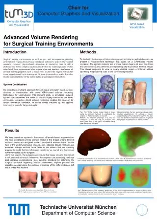
Chair for Computer Graphics and Visualization Computer Graphics GPU-based Visualization and Visualization Advanced Volume Rendering for Surgical Training Environments Introduction Surgical training environments as well as pre- and intra-operative planning environments require physics-based simulation systems to achieve the required accuracy. However, physics-based simulation at interactive rates is still a challenge due to the complex equations involved in the solution process. On the other hand, these environments often require a fast and intuitive visualization of internal material properties such as tissue stress to allow the surgeon to track the tissue stress induced by an intervention. If tissue is stressed too much, this often implies additional risks for the patient during a real surgical intervention. System Contribution By exploiting a multigrid approach for soft-tissue simulation based on finite elements in combination with novel GPU-based volume rendering techniques for unstructured finite-element grids, a simulation support system that addresses the needs in virtual surgery simulators has been developed. Interactive direct volume rendering enables the surgeon to obtain immediate feedback on tissue stress induced by the applied intervention even for large data-sets. Methods To deal with the barrage of information present in today’s medical datasets, we present a focus+context technique that builds on a GPU-based volume raycaster. The system extracts two or more feature layers (at least one focus and one context) and performs a compositing step to produce the final image. This allows surgeons to view a clearly defined region of interest without sacrificing the positional cues of the surrounding material. Top: The Visible Human (male) head is shown using two different methods to emphasize the location of the bone relative to the skin. Bottom: Two different renderings of a hand are compared. To the left, only iso-surfaces have been used, while to the right a volumetric tissue-layer (blue) was included. We demonstrate that our system produces highly intuitive visualizations. To achieve a natural understanding of the materials present in this data set, skin and bone shaders have been integrated to highlight the joint between femur and tibia. Results We have tested our system in the context of female breast augmentation. We have generated a finite-element model of the breast, where different stiffness values are assigned to each tetrahedral element based on the type of the underlying tissue (muscle, skin, adipose tissue). Implants are modelled through artificial force fields in the interior that are carefully adapted to model the kind of implant (anatomic vs. round) and the implant volume selected by the surgeon. Using our system, the surgeon can ensure that tissue close to the implant is not stressed too much. Moreover, the surgeon can potentially minimize post-operative complications (e.g., swelling, bleeding) by optimizing the surgical approach regarding implant parameters, implant position and operative access taking the material properties of the different breast soft tissue types into account. On the left, the boundary of an undeformed liver is shown. On the right, the deformed liver is visualized by means of direct volume rendering. The internal stress induced by the deformation is highlighted with green color. On the left, the surface scan of a female breast is shown. On the right, anatomic implants of size 150ml are simulated. Left: The outer surface of the volumetric model used for the physics-based simulation of gravity is shown. Right: Visualization of internal stress values. Red indicates high tissue stress while green marks undeformed regions. Such a visualization can assist in plastic and reconstructive surgery planning. Technische Universität München Department of Computer Science


