Download
1 / 44
460 likes | 549 Views
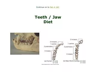
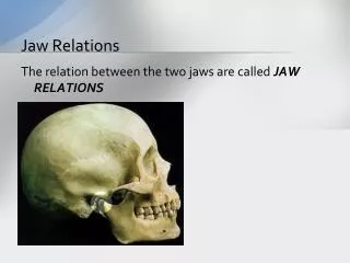
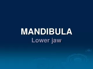
MANDIBULA Lower Jaw. Anatomy Clinical notes Dentoalveolar topography Nerve and blood supply. A single facial bone that is the only freely movable part of the skull During the development the mandible originates from two bones which unite together and form the

E N D
Anatomy • Clinical notes • Dentoalveolar topography • Nerve and blood supply
A single facial bone that is the only freely movable part of the skull During the development the mandible originates from two bones which unite together and form the lower jaw
Childhood • mandibular corpus is low • the body contains the sockets of deciduous teeth • the angle between corpus and ramus is 150° • mental foramen lies on the lower edge of corpus
Adulthood • the angle is much sharper – about 120° • condylar process is higher than the coronoid process and the sigmoid notch becomes deeper
Old age • after the loss of teeth, the body is reduced in volume (atrophy of the alveolar process) →mandibular foramen is closer to the alveolar border • enlargement of the angle to 160° • deepen pterygoid fovea → neck is tapered
Alveolar process • The portion of the jawbone that contains theteeth and the alveoli in which they are suspended • The development is dependent on tooth eruption and its maintenance on tooth retention • Is composed of compact bone (0.1-0.8 mm) that enclose the spongiosa
Alveolus Compact bone(labial cortical plate) Compact bone(lingual cortical plate) Spongy bone
Alveolus • Is composed of a thin plateofcortical bone with numerous perforations (or cribriform plate) that allow the passage of blood vesselsbetween the bone marrow spaces and the periodontal ligament • The coronal rim of the alveolar bone forms the alveolar crest, which generally parallels the cemento-enamel junction at a distance of 1-2 mm apical to it
Bundle bone = the inner portion of the bone of the alveolus that surrounds teeth and into which the collagen fibers of the periodontal ligament are embedded • Radiographically, thebundle bone is the laminadura
Interalveolar septum (spongy bone) 0.7-14 mm Alveolus (compact bone)
Resorption of alveolar bone Decreased bone (osteopenia) of alveolar process is noted when there is inactivity of tooth that does not have an antagonist
Reconstruction of alveolar bone • The whole life the bone keep the potential to reconstruction • Bone is resorbed on the side of pressure and opposed on the site of tension is regenerated • Movement of a tooth by extrusion involves applying traction forces in all regions of the periodontal ligament to stimulate marginal apposition of crestal bone
Lingual foramen (canal) The contents of theforamen (foramina) were foundto be an artery Median
Lateral (orifices accessories) CT • Inner area of mentum • Sup. and inf. retromental for. • Unilateral, bilateral or mutliple • In neighbourhood of mylohyoid line CAVE! Bleeding (implant placement)
Mandibular foramen • Beginning of canalis mandibulae • Inner surface of ramus mandibulae • Middleline between anterior and posterior edge of ramus • 1 cm above M3 • 2 cm behind M3 CAVE! Local anesthesy
Mandibular canal • Is placed under the alveoli and communicates with them by small openings • Contains the inferior alveolar nerve, artery, vein
Demarcate of the compact bone (noticeable to x-ray) • On arriving at the incisor teeth, it turns back to communicate with the mental foramen, giving off a small canal known as the mandibular incisive canal
Canalis mandib. bifidus Summary 0.9%
Mental foramen • The position of this foramen is most frequently near the apex of the mandibular second premolar and rested between the premolars • The foramen open upward and slightly posteriorly in adults • The foramen open straight upward in newborns
Incisive canal Summary 96% Demarcate of the compact bone (noticeable to x-ray)
Dentoalveolar topography Important foranesthesia, extraction, injury, implantology, endodontic treatment ... The transverse asymmetry of alveolus The rate of the spongy and the compact bone The relationship the roots the lower jaw to neighbouring structures
1. The transverse asymmetry of alveolus The dental and skeletal arch are asymmetric !
2. The rate of the spongy/compact bone • The layer of compact bone is thicker than in the upper jaw • Roots of the incisivi and canini teeth are surrounded by the compact bone • Roots of the premolars and molars are surrounded by the pre- and retroalveolar spongy bonethat is thin, fragible
Incisivi, Canini Compact bone only • CAVE! • Fractures by extraction ! • Root of the 3nd tooth – fracture of mandible !
Premolars Molars Compact bone and variable thickness of spongy bone bucally and lingually (linea mylohyoidea)
3. The relationship the roots the lower jaw to neighbouring structures Canalis mandibulae (incisivus, mentalis)
Canalis mandibulae • variable layer of spongy bone • dehiscence of the canal and the alv. Canalis incisivus • variable layer of spongy bone Canalis mentalis • variable layer of spongy bone • beneath the mandibular 4, 5
Trigeminal nerve Alveolar inferior nerve mental nerve incisive nerves Mylohyoid nerve Buccal nerve Lingual nerve
Variation Important for anesthesy ! 1.Mylohyoid nerve Can conveys impulses from the incisive, canineandpremolar teeth and gingiva !
2. Sometimes the branches entering separated bony channels laterocranial of mandible foramen and M3, M2 The nerves entering the mandible at the retromolar fossa
Maxillary artery Inferior alveolar arterymylohyoid a. dent. et interalveolar a. mental a. incisive a. Facial artery submental a. Lingual artery sublingual a.