Discovering the Veins: Locate and Understand One-Way Valves in Your Body
340 likes | 465 Views
Explore the fascinating world of your circulatory system by learning how to identify one-way valves in the veins of your arm. Our step-by-step guide allows you to understand blood flow direction and valve function through simple techniques. As you press and slide your thumb along the longest vein visible on your wrist, you'll uncover the mechanics of this crucial circulatory system component. Gain insights into the heart's pumping action and the roles of arteries, veins, and capillaries as you become a 'Valve Detective' in your own body!

Discovering the Veins: Locate and Understand One-Way Valves in Your Body
E N D
Presentation Transcript
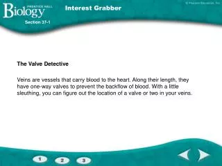
Interest Grabber Section 37-1 The Valve Detective • Veins are vessels that carry blood to the heart. Along their length, they have one-way valves to prevent the backflow of blood. With a little sleuthing, you can figure out the location of a valve or two in your veins.
Interest Grabber continued Section 37-1 • 1. Choose the longest vein you can see on the inner side of your wrist. Starting as close to your wrist as possible, press your thumb on the vein and slide it along the vein up your arm. Did the length of the vein remain blue? • 2. Repeat this process, but in the opposite direction, moving your thumb along the vein from the far end to the end closest to your wrist. Did the length of the vein remain blue? • 3. In which direction is your blood flowing in this vein? How can you tell? Can you tell where a valve is located? Explain your answer.
Section Outline Section 37-1 • 37–1 The Circulatory System A. Functions of the Circulatory System B. The Heart 1. Circulation Through the Body 2. Blood Flow Through the Heart 3. Heartbeat C. Blood Vessels 1. Arteries 2. Capillaries 3. Veins D. Blood Pressure E. Disorders of the Circulatory System 1. High Blood Pressure 2. Heart Attack 3. Stroke 4. Prevention of Circulatory System Disorders
Structures and Functions of the Circulatory System • The human circulatory system consists of the heart, a series of blood vessels, and the blood that flows through them. • The circulatory system is responsible for transporting nutrients and waste products throughout the body.
The Heart • The heart is composed almost entirely of muscle. • Its job is to pump blood throughout the body. • Enclosed and protected by pericardium. • Myocardium is the muscle layer.
Pulmonary and Systemic Circulation • Pulmonary circulation refers to the blood vessels that carry blood from the hear to the lungs. • Systemic circulation refers to the blood vessels that carry blood from the heart to all other parts of the body.
The Sinoatrial Node Section 37-1 Contraction of Atria Contraction of Ventricles Sinoatrial (SA) node Conducting fibers Atrioventricular (AV) node
Capillaries of head and arms Superior vena cava Pulmonary artery Aorta Pulmonary vein Capillaries of left lung Capillaries of right lung Inferior vena cava Capillaries of abdominal organs and legs Figure 37-2 The Circulatory System Section 37-1
Aorta Brings oxygen-rich blood from the left ventricle to the rest of the body Superior Vena Cava Large vein that brings oxygen-poor blood from the upper part of the body to the right atrium Pulmonary Arteries Bring oxygen-poor blood to the lungs Pulmonary Veins Bring oxygen-rich blood from each of the lungs to the left atrium Pulmonary Valve Prevents blood from flowing back into the right ventricle after it has entered the pulmonary artery Aortic Valve Prevents blood from flowing back into the left ventricle after it has entered the aorta Mitral Valve Prevents blood from flowing back into the left atrium after it has entered the left ventricle Tricuspid Valve Prevents blood from flowing back into the right atrium after it has entered the right ventricle Inferior Vena Cava Vein that brings oxygen-poor blood from the lower part of the body to the right atrium Figure 37-3 The Structures of the Heart Section 37-1 Left Atrium Right Atrium Left Ventricle Septum Right Ventricle
Blood Vessels • Arteries carry blood away from the heart. • Veins carry blood toward the heart. • Capillaries are the smallest blood vessels. • Most nutrient and waste product transfer at the cellular level is done by capillaries.
Endothelium Arteriole Venule Connective tissue Connective tissue Smooth muscle Smooth muscle Endothelium Valve Endothelium Figure 37-5 The Three Types of Blood Vessels Section 37-1 Vein Artery Capillary
Blood Pressure • Blood pressure is measured with a sphygmomanometer. • First number is systolic pressure; the force felt in the arteries when the ventricles contract. • Second number is diastolic pressure; the force felt in the arteries when the ventricles relax. • Typical blood pressure is 120/80.
Interest Grabber Section 37-2 Designer Blood • The federal government wants to find ways to make the blood supply safer for everyone who needs blood. However, no one has yet found a way to find and eliminate all disease-causing agents in the blood. Imagine that you are the head of a biotechnology company and think that you can design a safe alternative — artificial blood.
Interest Grabber continued Section 37-2 • 1. What characteristics would artificial blood need to take the place of real blood? • 2. Do you think that artificial blood could completely replace real blood? Explain your answer.
Section Outline Section 37-2 • 37–2 Blood and the Lymphatic System A. Blood Plasma B. Blood Cells 1. Red Blood Cells 2. White Blood Cells 3. Platelets and Blood Clotting C. The Lymphatic System
Blood • Blood is 8% of your total mass. • 45% of blood is composed of cells. • 55% of blood is plasma. • Plasma is 90% water and 10% gases, salts, proteins, nutrients, enzymes, hormones, and waste products. • Plasma proteins: • Albumins regulate pressure and volume. • Globulins fight infection. • Fibrinogens help clotting.
Blood Cells • Red blood cells (erythrocytes) transport oxygen. • Hemoglobin allows this by binding to oxygen. • White blood cells (leukocytes) guard against infection, fight parasites, and attack bacteria. • Blood clotting is made possible byby plasma proteins and cell fragments called platelets.
Blood Transfusions Section 37-2 Blood Type of Recipient Blood Type of Donor A B AB O A B AB O Unsuccessful transfusion Successful transfusion
Figure 37-7 Blood Section 37-2 Plasma Platelets White blood cells Red blood cells Whole Blood Sample Sample Placed in Centrifuge Blood Sample That Has Been Centrifuged
Figure 37-7 Blood Section 37-2 Plasma Platelets White blood cells Red blood cells Whole Blood Sample Sample Placed in Centrifuge Blood Sample That Has Been Centrifuged
Figure 37-7 Blood Section 37-2 Plasma Platelets White blood cells Red blood cells Whole Blood Sample Sample Placed in Centrifuge Blood Sample That Has Been Centrifuged
Figure 37-9 Types of White Blood Cells Section 37-2 Cell Type Neutrophils Eosinophils Basophils Monocytes Lymphocytes Function Engulf and destroy small bacteria and foreign substances Attack parasites; limit inflammation associated with allergic reactions Release histamines that cause inflammation; release anticoagulants, which prevent blood clots Give rise to leukocytes that engulf and destroy large bacteria and substances Some destroy foreign cells by causing their membranes to rupture; some develop into cells that produce antibodies, which target specific foreign substances
Figure 37-10 Blood Clotting Section 37-2 Break in Capillary Wall Blood vessels injured. Clumping of Platelets Platelets clump at the site and release thromboplastin. Thromboplastin converts prothrombin into thrombin.. Clot Forms Thrombin converts fibrinogen into fibrin, which causes a clot. The clot prevents further loss of blood..
Lymphatic System • A network of vessels, nodes, and organs called the lymphatic system collects the fluid that is lost by the blood and returns it back to the circulatory system.
Figure 37-12 The Lymphatic System Section 37-2 Superior vena cava Thymus Heart Thoracic duct Spleen Lymph nodes Lymph vessels
Interest Grabber Section 37-3 Hold That Breath! • Do not perform this activity if you have any breathing problems. Working with a partner, count the number of breaths you take in 15 seconds. Multiply that number by 4 for the number of breaths per minute. Your partner will act as the timer/recorder. Repeat the procedure three times and take an average. Now, take a deep breath and hold it for as long as you can. Have your partner record your time. Repeat the procedure three times and take an average. Switch roles with your partner and repeat the procedure. Exchange data with other groups and answer the following questions.
Interest Grabber continued Section 37-3 • 1. What was the range of breathing rates? • 2. Why are there differences in breathing rates among members of the class? • 3. What was the average length of time classmates could hold their breath? • 4. What factors might affect how long you could hold your breath? • 5. A child having a tantrum declares she is going to hold her breath “until I turn blue!” Do you think this is possible? Explain your answer.
Section Outline Section 37-3 • 37–3 The Respiratory System A. What Is Respiration? B. The Human Respiratory System C. Gas Exchange D. Breathing E. How Breathing Is Controlled F. Tobacco and the Respiratory System 1. Substances in Tobacco 2. Diseases Caused by Smoking 3. Smoking and the Nonsmoker 4. Dealing With Tobacco
Respiratory System • The basic function of the respiratory system is to bring about the exchange of oxygen and carbon dioxide between the blood, air, and tissues.
Figure 37-14 The Respiratory System Section 37-3 Pharynx Larynx Nose Trachea Mouth Lung Bronchiole Bronchus Epiglottis Alveoli Bronchioles Diaphragm Capillaries Edge of pleural membrane
Figure 37-15 Gas Exchange in the Lungs Section 37-3 Alveoli Bronchiole Capillary
Figure 37-16 The Mechanics of Breathing Section 37-3 Air exhaled Air inhaled Rib cage descends Rib cage rises Diaphragm Diaphragm Inhalation Exhalation
Figure 37-16 The Mechanics of Breathing Section 37-3 Air exhaled Air inhaled Rib cage descends Rib cage rises Diaphragm Diaphragm Inhalation Exhalation
Flowchart Section 37-3 Movement of Oxygen and Carbon Dioxide In and Out of the Respiratory System Nasal cavities Oxygen-rich air from environment Pharynx Trachea Bronchi Oxygen and carbon dioxide exchange at alveoli Bronchi Bronchioles Bronchioles Alveoli Carbon dioxide-rich air to the environment Nasal cavities Pharynx Trachea