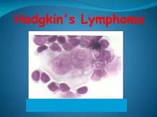
Lymphoma
Lymphoma. Uglyar T.Y. Adapted from Joe Waller, MD 2013 Drs. Wang and Young and By David Lee MD, FRCPC. Conceptualizing lymphoma. neoplasms of lymphoid origin, typically causing lymphadenopathy leukemia vs lymphoma lymphomas as clonal expansions of cells at certain developmental stages. ALL.


Lymphoma
E N D
Presentation Transcript
Lymphoma Uglyar T.Y. Adapted from Joe Waller, MD 2013 Drs. Wang and Young and By David Lee MD, FRCPC
Conceptualizing lymphoma • neoplasms of lymphoid origin, typically causing lymphadenopathy • leukemia vs lymphoma • lymphomas as clonal expansions of cells at certain developmental stages
ALL CLL Lymphomas MM Neutrophils AML Myeloproliferative disorders Myeloid progenitor Eosinophils Hematopoietic stem cell Basophils Monocytes Platelets Red cells naïve germinal center B-lymphocytes Plasma cells Lymphoid progenitor T-lymphocytes
CLL MM ALL DLBCL, FL, HL B-cell development memory B-cell germinal center B-cell stem cell mature naive B-cell lymphoid progenitor progenitor-B pre-B immature B-cell plasma cell Bone marrow Lymphoid tissue
Risk Factors • • Mostly unknown, although both genetic and infectious • processes are suspected • • Living in Western countries, being of higher social class, • more educated. • • Genetic pre-disposition, clusters noted in siblings with • similar HLA genotypes. • – Mack et al: 99x risk in monozygotic vs dizygotic twins • • EBV (MC subtype) • • HIV+ pts have different patterns of spread, more • systemic sx, poor tolerance to chemo • • Children do better than adults
Category Survival of untreated patients Curability To treat or not to treat Non-Hodgkin lymphoma Indolent Years Generally not curable Generally defer Rx if asymptomatic Aggressive Months Curable in some Treat Very aggressive Weeks Curable in some Treat Hodgkin lymphoma All types Variable – months to years Curable in most Treat A practical way to think of lymphoma
Staging • Stage I: a single LN region (on either side of the diaphragm) • Stage II: two or more LN regions of the same side of the diaphragm • Stage III: both sides of the diaphragm • Stage III-1: upper abd: splenic, celiac, portal LN only, <4 splenic • nodules • Stage III-2: lower abd: Paraaortic, mesenteric, pelvic • Stage III(S)+ Minimal: <4 splenic nodules • Stage III(S)+ Extensive: 5 or more splenic nodules • Stage IV: diffuse involvement of extralymphatic tissues, with or • without simultaneous LN involvement. • E subtypes: extranodal disease • S subtype: spleen involvement • “A” and “B”: absent or present “B” symptoms. • X subtype: bulky disease of > 1/3 thoracic diameter or > 10 cm
International Prognostic Score • In patients with Stage III-IV disease, each of the following factors • reduces survival by 7%: • Age >45 • Male sex • Stage IV disease • Albumin < 4g/dL • Hb<10.5 • WBC>15,000 • Lymphoctes count <8% or ALC<600 • Used for individualized treatment management
Other Manifestations • SVC syndrome • • Spinal Cord • Compression • • Bone involvement • • Hepatic involvement • • Renal involvement • • Infections • • Immunologic • Abnormalities • • Rarely: • – Waldeyer's Ring, • Peyer's patches, CNS, • skin
Epidemiology of lymphomas • 5th most frequently diagnosed cancer in both sexes • males > females • incidence • NHL increasing • Hodgkin lymphoma stable • Epidemiology • • ~8000 new cases of Hodgkin’s Disease in • the U.S. in 2008, causing ~1500 deaths • • M:F ratio is 1.3:1; more pronounced in • children • • Bimodal age distribution: 2-3rd decade, • and 6-7th decade.
Clinical manifestations • Variable • severity: asymptomatic to extremely ill • time course: evolution over weeks, months, or years • Systemic manifestations • fever, night sweats, weight loss, anorexia, pruritis • Local manifestations • lymphadenopathy, splenomegaly most common • any tissue potentially can be infiltrated
complications of lymphoma • bone marrow failure (infiltration) • CNS infiltration • immune hemolysis or thrombocytopenia • compression of structures (eg spinal cord, ureters) • pleural/pericardial effusions, ascites
Diagnosis requires an adequate biopsy Work Up • Diagnosis should be biopsy-proven before treatment is initiated • Need enough tissue to assess cells and architecture • open bx vs core needle bx vs FNA • Excisional biopsy • – Most commonly of cervical nodes • – Presence of RS cells is necessary but not sufficient • • Laparotomy • – 1960’s • – Determine extent of disease below diaphragm • – Largely eliminated by more effective chemo regimens • – EORTC study did not show survival benefit for • pathologic staging over clinical staging (Carde et al. • JCO 1993)
Adverse Prognostic Factors • • B symptoms esp. weight loss and night sweats. • • Pruritis • • Higher stage, esp.with bone marrow or organ • involvement. • • Bulky disease with large tumor burden. This includes • large mediastinal lymphadenopathy, which is >1/3 of • maximal thoracic diameter (T5-T6). • • Worrisome labs include ESR>70 and high serum copper. • • Older age • • LD type • • male
CHOP Chemotherapy • Cyclophosphamide (Cytoxan) • Hydroxydaunorubicin (Adriamycin) • Oncovin (vincristine) • Prednisone
Follicular lymphoma • most common type of “indolent” lymphoma • usually widespread at presentation • often asymptomatic • not curable (some exceptions) • associated with BCL-2 gene rearrangement [t(14;18)] • cell of origin: germinal center B-cell
defer treatment if asymptomatic (“watch-and-wait”) • several chemotherapy options if symptomatic • median survival: years • despite “indolent” label, morbidity and mortality can be considerable • transformation to aggressive lymphoma can occur
Diffuse large B-cell lymphoma • most common type of “aggressive” lymphoma • usually symptomatic • extranodal involvement is common • cell of origin: germinal center B-cell • treatment should be offered • curable in ~ 40%
Treatment Options:Aggressive Lymphomas Aggressive • Diffuse large cell lymphoma, large cell anaplastic lymphoma, peripheral T cell lymphoma. Very Aggressive • Burkitt’s lymphoma and lymphoblastic lymphoma.
Treatment Options for Advanced Stage Aggressive Lymphomas • Systemic chemotherapy • CHOP (± Rituxan for over 70 age group) • ± Intrathecal chemotherapy • AIDS patients and CNS involvement • ± Radiotherapy • Spinal cord compression, bulky disease
Burkitt’s Lymphoma • African variety: jaw tumor, strongly linked to Epstein-Barr Virus infection. • In U.S., about 50% EBV infection. • May present as abdominal mass. • Most rapidly growing human tumor. • Typical chromosome abnormality: c-myc oncogene linked to one of the immunoglobulin genes.
Burkitt’s Lymphoma • Treated with multidrug regimen similar to pediatric leukemia/lymphoma regimens. • Treated with multidrug regimen similar to pediatric leukemia/lymphoma regimens.
MALT Lymphoma • Mucosa-Associated Lymphoid Tissue • Chronic infection of the stomach by Helicobacter pylori. • Localized to the stomach, indolent course. • Can be cured in many cases by antibiotics against H. pylori.
Treatment Options for Early Stage Aggressive Lymphomas • Often in Stage I or II • potentially curable • disseminates through bloodstream early • must use systemic chemotherapy • CHOP x 6 cycles • CHOP x 3 cycles followed by radiotherapy
Hodgkin lymphoma Thomas Hodgkin (1798-1866)
Classical Hodgkin Lymphoma Hodgkin’s Disease
Hodgkin’s Disease • One-seventh as common a snon-Hodgkin’s lymphoma. • Highly treatable and curable, even when disseminated. • Presence of Reed-Sternberg cell is necessary to make diagnosis.
Subtypes of Hodgkin’s Disease • Lymphocyte predominant • Nodular sclerosis • Mixed cellularity • Lymphocyte depleted • Unlike non-Hodgkin’s lymphoma, in Hodgkin’s Disease • the histologic subtype does not determine how the • disease is treated.
CHL Pathologic Variants • Nodular Sclerosis (NS) (70%) • • Large RS cells • • Cervical nodes • • Anterior mediastinum • • Adolescent patients • • Lymphocyte Rich (5%) • • Rare RS cells. Many lymphocytes. Age <35 y/o with localized disease. Good • prognosis. M>F (4:1). • • Lymphocyte Depleted (rare) • • Many RS cells, few lymphocytes • • Age > 50. • • Diffuse abdominal disease, marrow, and liver involvement. Most patients p/w • advanced disease • • Poorest prognosis • • Mixed cellularity (25%) • • Moderate RS cells, mixed infiltrate of neutrophils, eosinophils, and plasma cells. • • Age 30-50, EBV associated, developing countries • • Retro-peritoneal presentation • • Intermediate prognosis
Hodgkin lymphoma • cell of origin: germinal centre B-cell • Reed-Sternberg cells (or RS variants) in the affected tissues • most cells in affected lymph node are polyclonal reactive lymphoid cells, not neoplastic cells
Histology • Reed-Stenberg Cell: “owl eyes” • – Large, with abundant cytoplasm, 2-3 nuclei with • prominent nucleolus “owl eyes” appearance • – NOT pathognomonic, can be reactive, infectious or • malignant • – RS cells stain for CD30/15+, but are CD45/20- • *Contrast w/ NLPHL which are CD30/15-, • CD45/20+
RS cell and variants classic RS cell lacunar cell popcorn cell (lymphocyte predominance) (mixed cellularity) (nodular sclerosis)
Molecular Biology • The Reed Sternberg (RS) cell likely arises from • either lymphocyte or antigen-presenting cells of • the monocyte-macrophage line. Regarding • lymphocyte origin, 60% of RS cells have T or B • cell specific antigens, and B cells are the usual • target for EBV
Molecular Biology • RS-like cells are found in several infectious, inflammatory, and • neoplastic conditions including infectious mononucleosis, reactive • lymphoid hyperplasia, and immunoblastic lymphoma. • • Thus, diagnosing Hodgkin’s depends on finding RS cells in the • appropriate clinical setting. The lymphocytes are predominantly CD- • 4 positive T-cells. • • The BCL2 Oncogene is found in 1/3 of Hodgkins patients. • • The p53 suppressor gene is found in almost all Hodgkin’s patients • except those with lymphocyte predominant disease. • • The common t(14:18) translocation of B cell lymphoma are RARE in • RS cells.
A possible model of pathogenesis loss of apoptosis transforming event(s) EBV? cytokines germinal centre B cell RS cell inflammatory response
Etiology ofHodgkin’s Disease • Reed-Sternberg cells are the malignant cells. • Minor population in the malignant tissues • many normal lymphocytes, eosinophils, other cells • Cell of origin is unknown: T, B, both, neither. • Some R-S cells contain EBV genomes.
Epidemiology • less frequent than non-Hodgkin lymphoma • overall M>F • peak incidence in 3rd decade
Clinical Features • T cell mediated immune deficiency, even in early stage disease. Prone to infections: • Herpes zoster (“shingles”) in one fourth of patients • Fungal or mycobacterial infections • Immune defect may persist even after lymphoma is cured.
Clinical manifestations: • lymphadenopathy • contiguous spread • extranodal sites relatively uncommon except in advanced disease • “B” symptoms
Clinical Features • Predictable contiguous spread of disease: • cervical nodes to mediastinum or axilla • mediastinum to periaortic nodes or spleen, etc. • Basis for staging and treatment decisions.
Diagnosis • Excisional biopsy of a lymph node. Fine needle aspirate is not sufficient to make the diagnosis of Hodgkin’s disease.
Staging of Hodgkin’s Disease Same as for non-Hodgkin’s: H + P, labs, CT scans, bone marrow biopsy PLUS: Gallium scan Lymphangiogram or staging laparotomy ONLY if results would affect treatment decisions
Chemotherapy Regimens MOPP Mechlorethamine, Oncovin, Procarbazine, Prednisone ABVD Adriamycin, Bleomycin, Vinblastine, Dacarbazine BEACOPP
Treatment Options Often, patients who relapse after radiotherapy can be cured by salvage chemotherapy. Combined chemotherapy and radiotherapy is given for bulky mediastinal masses. Chemotherapy now being tested for earlier stages of the disease.
Late Complications ofHodgkin’s Disease High incidence of second malignancies leukemia first 10 years, solid tumors over time. Leukemia in patients receiving alkylating agents or combined chemo/XRT. Lung cancer and breast cancer in patients receiving XRT to chest. Lung cancer especially high in smokers.
Late Complications ofHodgkin’s Disease Hypothyroidism after irradiation of the neck. Constrictive pericarditis after radiotherapy to the mediastinum. Infertility after use of alkylating agents. Heart failure after Adriamycin treatment.