Download
1 / 81
820 likes | 1.28k Views
Intensive Care After Neurosurgery JEAN-LOUIS VINCENT(41) Miller(94). Saeed Abbasi , MD, FCCM. Intensive Care After Neurosurgery. Overview Prevention and Management of Systemic Complications After Neurosurgery Prevention and Management of Neurosurgical Postoperative Complications

E N D
Intensive Care After NeurosurgeryJEAN-LOUIS VINCENT(41)Miller(94) SaeedAbbasi, MD, FCCM
Intensive Care After Neurosurgery • Overview • Prevention and Management of Systemic Complications After Neurosurgery • Prevention and Management of Neurosurgical Postoperative Complications • Admission Examination and Monitoring in the Intensive Care Unit • Systemic Monitoring: Cardiopulmonary, Respiratory Status, and Temperature • Brain Monitoring and Specific Therapeutic Approaches • Neuroprotection
Overview • Collaboration between various specialists: neurosurgeons, intensivists, and neuroradiologists • Admission policy
Priorities and Goals of Postoperative Neurosurgical Care • Early detection and treatment of postsurgical complications • Preventing second insults
Postoperative complications may be systemic or neurosurgical
Prevention and Management of SystemicComplications After Neurosurgery
GENERAL PRINCIPLES AND SECOND INSULTS • Follows general principles of “intensive care” medicine • Systemic complications and second insults may initiate or aggravate cerebral damage • Conversely, CNS events may induce systemic derangement : response to raised intracranial pressure (ICP)
GENERAL PRINCIPLES AND SECOND INSULTS • Many drugs routinely used in neurosurgical patients may cause complications or side effects • steroids • antiepileptic agents • Spinal cord injury : loss of autonomic sympathetic function
CARDIAC DYSFUNCTION • Electrocardiographic (ECG) abnormalities : diffuse ST-segment changes mimicking cardiac ischemia and cardiac arrhythmias, may be caused by SAH, TBI, or raised ICP • Takotsubo syndrome : The left ventricle suffers a typical bulging (indicating ischemic changes and functional impairment)
NEUROGENIC PULMONARY EDEMA • After a variety of neurosurgical procedures : brain tumors (particularly those resected in the posterior fossa), cysts, hydrocephalus, intracranial hemorrhages, and brainstem lesions • 9% mortality rate • Initial 4 hours after the neurologic event • More common in women than in men
NEUROGENIC PULMONARY EDEMA • Mechanisms unclear • Sudden central sympathetic discharge may trigger pulmonary venoconstriction, systemic arterial hypertension, increased left ventricle afterload, increased capillary permeability in the pulmonary vascular bed, and simultaneously cause cardiac ischemia and ventricular failure
NEUROGENIC PULMONARY EDEMA • Therapeutic measures : • Supportive • Opioids and sedatives • Supplemental oxygen • Tracheal intubation with mechanical ventilation and application of PEEP in 75% of patients • Diuretics • Vasoactive drugs
HYPERCOAGULOPATHY AND THROMBOSISPROPHYLAXIS • DVT : 18% to 50% • PE in 0% to 25% • Mechanical therapies carry less associated risk, but pharmacologic approaches are more effective
HYPERCOAGULOPATHY AND THROMBOSISPROPHYLAXIS • Overall, existing evidence, however, shows that the beneficial effects in reducing DVT and in particular PE outweigh a slightly increased risk of clinically significant hemorrhagic complications with anticoagulant prophylaxis
Prevention and Management of Neurosurgical Postoperative Complications
SUPRATENTORIAL PROCEDURES • Postoperative Subgaleal Hematoma • In up to 11% • Can be minimized by routine use of postoperative wound drainage for 24 hours • Reoperation is seldom necessary
SUPRATENTORIAL PROCEDURES • Intracranial Hemorrhage • 1% of procedures • Intraparenchymal hematomas (43%-60%), epidural hematomas (28%-33%) and subdural hematomas (5%-7%) • Parenchymal hemorrhages • Most frequent • Generally occur at the site of operation • In rare cases, distant from site of operation • Should be considered in all patients who are not fully alert post anesthesia, as well as in those who exhibit secondary deterioration
SUPRATENTORIAL PROCEDURES • Postoperative Brain Swelling • Predisposing factors • Hypercapnia • Arterial hypertension • Hyponatremia • Obstruction of venous drainage • Silent or overt seizures during surgery or in the immediate postoperative phase • Brain swelling due to vasodilation : hyperventilation and barbiturate administration • Brain swelling due to cerebral edema : mild hyperventilation and osmotic agents
SUPRATENTORIAL PROCEDURES • Tension Pneumocephalus • Rewarming of air in the intracranial compartment postoperatively or continuous air leakage due to a cerebrospinal fluid (CSF) fistula of the skull base • Clinical symptomatology : decreasing level of consciousness, signs of raised ICP, and occasionally seizures • Generally self-limiting and do not require specific treatment.
SUPRATENTORIAL PROCEDURES • Seizures • Occult seizure activity can occur in 15% to 18% of patients with moderate and severe TBI • Prophylactic antiseizure indications are restricted to patients with a higher risk: • Cerebrovascular surgery (arteriovenous malformation, aneurysm) • Cerebral abscess and subdural empyema • Convexity and parafalcialmeningiomas • Penetrating brain injury • Compound depressed skull fracture • some centers recommending a treatment duration of 2 weeks and others continuing for at least 3 months • In any case of unexplained neurologic deterioration or delayed awakening from anesthesia, the possibility of seizures should be considered
INFRATENTORIAL PROCEDURES • Rapid deterioration because of the relatively small infratentorial volume reserve and the immediate compression of the brainstem • Irritation of the brainstem : large swings in arterial BP • Lesions of the lower cranial nerves : diminished gag reflex, with increased risk of aspiration and pneumonia
INFRATENTORIAL PROCEDURES • After any infratentorial procedure, the risk of acute hydrocephalus due to obstruction at the level of the fourth ventricle is present • Routine admission of all patients who have undergone posterior fossa surgery to the ICU • Particular attention should be paid to the presence of the gag reflex before extubation and in the early stages after extubation
INFRATENTORIAL PROCEDURES • Aseptic meningitis • Meningeal symptoms, headaches, and an inflammatory response of the CSF in the absence of evidence for infection • The origin of this syndrome has not been fully clarified
CEREBROVASCULAR PROCEDURES • The main cerebral complications are: 1. Rebleeding 2. Delayed cerebral ischemia 3. Hydrocephalus
CEREBROVASCULAR PROCEDURES • Rebleeding • first weeks after the aneurysmal rupture • Delayed cerebral ischemia (DCI) • Angiographic vasospasm : 67% of untreated patients • The time of maximum spasm around the end of the first week • DCI cannot always be attributed to vasospasm but more to the occurrence of microthrombosis • Oral calcium antagonists in preventing delayed ischemic deficits • Triple-H therapy (hypervolemia, hypertension, and hemodilution)
CEREBROVASCULAR PROCEDURES • Hydrocephalus • Not uncommon • Spontaneous improvement of hydrocephalus has been reported in approximately half of patients
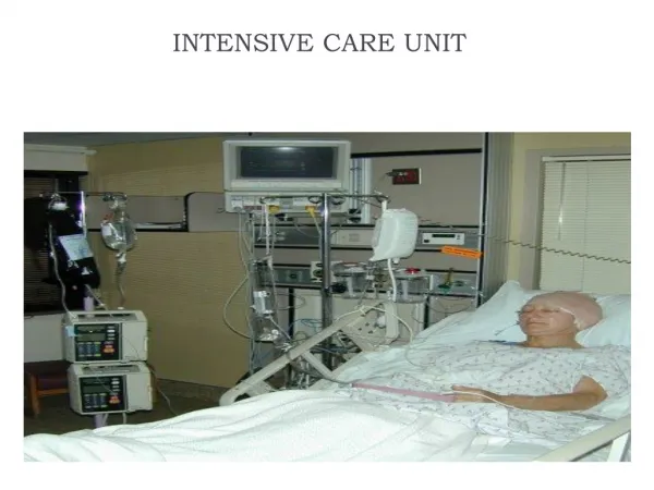
Admission Examination and Monitoring in the Intensive Care Unit
EARLY EVALUATION • Glasgow Coma Scale • Pressure on the nail bed and supraorbital pressure
EARLY EVALUATION • The development of pupillary abnormalities is a sensitive indicator for pressure on the midbrain (tentorialherniation)
FURTHER EVALUATION • Evaluation is important, since cranial nerve deficits can require immediate treatment • Protection of the ocular bulb to prevent keratitis • Avoidance of oral feeding if swallowing is impaired
Systemic Monitoring: Cardiopulmonary,Respiratory Status, and Temperature • Invasive arterial BP monitoring is recommended • Hypovolemic shock • Skin pallor and poor capillary refill may precede a drop in BP • Hematocrit of approximately 30% to 33% as optimal in the acute postoperative period in patients in the neurosurgical ICU • After intracranial or spinal cord procedures aiming at a hemoglobin of at least 9-10 mg/dL
Systemic Monitoring: Cardiopulmonary,Respiratory Status, and Temperature • Cardiogenic shock: • Elderly patient • Takotsubo syndrome • Require sequential echocardiographic follow-up • Large pulmonary emboli, sepsis, or spinal paraplegia should also be considered in patients with systemic hypotension
Systemic Monitoring: Cardiopulmonary,Respiratory Status, and Temperature • Spinal distributive shock : • Hypotension is associated with bradycardia, with a pulse in the range of 35 to 50 • Should not be managed with excessive volume resuscitation but rather with vasopressors to restore α-adrenergic peripheral vasomotor tone
Systemic Monitoring: Cardiopulmonary,Respiratory Status, and Temperature • The combination of hypertension and bradycardia (Cushing response) • Potential of an expanding intracranial lesion and risk of brainstem herniation • Antihypertensive agents is contraindicated, and therapy should be aimed at the raised ICP
Systemic Monitoring: Cardiopulmonary,Respiratory Status, and Temperature • Core temperature should be kept lower than 38.0°C, using medications (e.g., acetaminophen, paracetamol, diclofenac) and surface or intravascular cooling • Hypothermia may be due to adrenal or pituitary insufficiency, hypothalamic disorders, hypoglycemia, or intraoperative exposure
Systemic Monitoring: Cardiopulmonary,Respiratory Status, and Temperature • Hypothermia complications : • Cardiovascular instability (mainly arrhythmias) • Coagulopathy • Electrolyte shifts • Fluid overload • Increased risk of infection • Shivering
BIOCHEMICAL PARAMETERS: ELECTROLYTES,OSMOLARITY, AND BLOOD GLUCOSE • Keeping biochemical parameters within physiologic ranges is obviously desirable, but this apparently simple goal may require a lot of work
ELECTROLYTES AND OSMOLARITY • General recommendation is that serum osmolarity should be kept below 320 mOsm • Sudden episodes of diabetes insipidus are likely • Cerebral salt waisting • Fluid restriction for correction should generally be avoided; it is often better to administer hypertonic saline
GLUCOSE • In our opinion, the currently available evidence would not support the use of tight glucose control in neurointensive care
Brain Monitoring and SpecificTherapeutic Approaches • ICP and CPP monitoring • Cerebral oxygenation • Continuous EEG • Magnetic resonance spectroscopy
INTRACRANIAL PRESSURE AND CEREBRALPERFUSION PRESSURE • ICP monitoring • severe brain injury (GCS < 8) • Abnormalities on the initial CT scan • Normal admission CT scan if two or more of the following features are present: • Age older than 40 years • Unilateral or bilateral motor posturing • Systolic BP less than 90 mm Hg • Routine ICP monitoring is not generally indicated in patients with mild or moderate TBI
INTRACRANIAL PRESSURE AND CEREBRALPERFUSION PRESSURE • ICP monitoring is further indicated in poor-grade patients with aneurysmal SAH • It may be considered in patients with other intracranial disorders who are sedated and ventilated and in whom the risk of raised ICP is considered present (postoperative swelling, stroke, Reye syndrome)
INTRACRANIAL PRESSURE AND CEREBRALPERFUSION PRESSURE • ICP monitoring carries a 0.5% risk of hemorrhage and a 2% risk of infection • Intraventricular catheters are preferable because they are accurate, can be recalibrated, and allow drainage of CSF • Intraparenchymal probes are user friendly and accurate • Less accurate data are provided by subdural catheters, and epidural probes are unreliable
INTRACRANIAL PRESSURE AND CEREBRALPERFUSION PRESSURE • Normal values for ICP are up to 15 mm Hg in adults, and consensus supports maintaining ICP below 20 mm Hg • More important is the trend over time and the relation to the arterial BP • MABP − ICP = CPP
TREATMENT OF CEREBRAL HERNIATION AND ELEVATED ICP • The emergency measures to be taken include : • Ventricular CSF drainage (if access available) • Bolus administration of high-dos hyperosmolar agents: mannitol: 1 to 1.5 g/kg bodyweight; hypertonic saline (HTS) 1 to 2 mL/ kg body weight 7.5% saline infused over 5 minutes • Rapid-sequence intubation and moderate hyperventilation
TREATMENT OF CEREBRAL HERNIATION AND ELEVATED ICP • Conservative therapy of elevated ICP includes: • Sedation, analgesia, and mild to moderate hyperventilation (30-35 mm Hg) • Osmotic therapy: preferably mannitol given in bolus infusions (dose: 0.25-0.5 g/kg bodyweight, or as indicated by monitoring). • Alternatively, HTS may be considered. Effective doses as bolus infusion range between 1 and 2 mL/kg of 7.5% saline. Effective doses as a continuous infusion of 3% range between 75 and 150 mL/h.
TREATMENT OF CEREBRAL HERNIATION AND ELEVATED ICP • CSF fluid drainage • Volume expansion and inotropes or vasopressors when arterial BP is insufficient to maintain CPP and CBF in a normovolemic patient