Download
1 / 35
360 likes | 554 Views
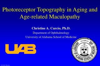
Photoreceptor Topography in Aging and Age-related Maculopathy. Christine A. Curcio, Ph.D. Department of Ophthalmology University of Alabama School of Medicine. Outline. Macula: cells and layers Photoreceptors as bioassay Aging & ARM: photoreceptor mosaic Aging & ARM: photoreceptor function

E N D
Photoreceptor Topography in Aging and Age-related Maculopathy Christine A. Curcio, Ph.D. Department of Ophthalmology University of Alabama School of Medicine
Outline • Macula: cells and layers • Photoreceptors as bioassay • Aging & ARM: photoreceptor mosaic • Aging & ARM: photoreceptor function • Possible explanations • Retinoid deficiency hypothesis • Implications for basic and clinical research
References • Curcio CA, Owsley C, Jackson GR: Spare the rods, save in the cones in aging and age-related maculopathy. Invest Ophthalmol Vis Sci 2000, 41:2015-2018 • Curcio CA: Photoreceptor topography in ageing and age-related maculopathy. Eye 2001, 15:376-383. • Jackson GR, Owsley C, Curcio CA: Photoreceptor degeneration and dysfunction in aging and age-related maculopathy. Ageing Research Reviews 2002, 1:381-396
Relative Rate of Rod and Cone Degeneration • Fundamental to each photoreceptor degeneration • Requires similar measures of rods and cones at same retinal locations in well-characterized eyes • Possible measures include numbers, morphology, imaging, and function
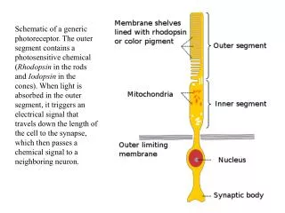
Why Study Photoreceptor Health? • Vision loss in ARM is due to dysfunction, death of photoreceptors • RPE/ Bruch’s membrane complex is vital to photoreceptors but difficult to study in vivo • Photoreceptor health is a direct bioassay of RPE/Bruch’s membrane health • Progress has been facilitated • Better understanding of dark adaptation • Grading systems for characterizing maculopathy
Photoreceptor Mosaic Curcio, Sloan, Kalina, Hendrickson. J Comp Neurol 1990, 292:497
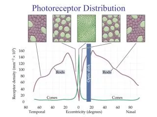
Cones Rods Macula: Photoreceptor Topography Anatomical and epidemiologic macula: 6 mm (21°) diameter Small, cone-dominated fovea Large, rod-dominated parafovea Curcio, Sloan, Kalina, Hendrickson. J Comp Neurol 1990, 292:497
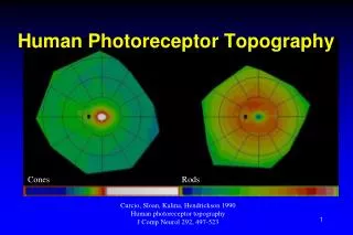
Human Photoreceptor Topography Curcio, Sloan, Kalina, Hendrickson. J Comp Neurol 1990, 292:497
Aging: Fovea & Parafovea Curcio. Eye 2001, 15:376
27-37 yr 82-90 yr Difference Topography of Age-Related Rod Loss Curcio, Millican, Allen, Kalina. IOVS 1993, 34:3278
Topography of Age-related Rod Loss (2) Curcio. Eye 2001, 15:376
Photoreceptors in ARM • 12 pairs of ARM eyes, donors 64-95 yr • 6 non-exudative (early and late) • 6 exudative • Photoreceptor counted in whole mounts • Loss relative to controls at each location • % of locations where rod loss>cone loss • Histopathology, histochemistry in fellow eye • Review of clinical records Curcio, Medeiros, Millican. IOVS 1996, 37:1236 Medeiros, Curcio. IOVS 2001, 42:795
Early ARM and Photoreceptors Curcio. Eye 2001, 15:376
Photoreceptor Loss & Fundus Adapted from Curcio, Medeiros, Millican. IOVS 1996, 37:1236
ExudativeARM &Photoreceptors Curcio. Eye 2001, 15:376
Rod and Cone Loss in ARM Rod loss > Cone loss 4/6 NE-ARM eyes 5/6 Ex-ARM eyes Curcio, Medeiros, Millican. IOVS 1996, 37:1236 Medeiros, Curcio. IOVS 2001, 42:795
Apoptotic Photoreceptors in ARM are Rods Dunaief, Dentchev, Ying, Milam Arch Ophthalmol 2002, 120:1435.
Support from Functional Studies • Large studies (99 adults, 80 early ARM patients) • Objectively characterized macular health • Rod and cone sensitivity at same retinal locations • Decrease throughout adulthood • Rod loss > cone loss in 80% of normal subjects • Declines further in early ARM, especially near fovea • Rod loss > cone loss in 87% of patients Aging: Jackson & Owsley. Vision Res. 2000;40:2467-2473. ARM: Owsley et al. IOVS 2000;41:267-273.
Aging:Scotopic Loss >Photopic Loss Jackson & Owsley. Vision Research 2000, 40:2467
Early ARM: Scotopic Loss > Photopic Loss Owsley, Jackson, Cideciyan, Huang, Fine, Ho, Maguire, Lolley, Jacobson. IOVS 2000, 41:267-273
Aging: Slower Dark Adaptation Jackson, Owsley, McGwin. Vision Res 1999, 39:3975
Early ARM: Slower Dark Adaptation Owsley, Jackson, White, Feist, Edwards. Ophthalmology 2001, 108:1196
Poor Scotopic Sensitivity/ Night Driving From Scilley, Jackson, Cideciyan, Maguire, Jacobson, Owsley. Ophthalmology 2002, 109:1235
Autofluorescence due to lipofuscin Human RPE Macular pigment, macaque (from Snodderly) Topographyof Effects Jackson, Owsley, Curcio. Ageing Research Reviews 2002, 1:381
Summary • Slowing of rod-mediated dark adaptation • Qualitative similarity of aging and ARM effects on photoreceptor function • Earlier loss of rods relative to cones • Topographic correspondence of dysfunction and loss to RPE/ Bruch’s pathology
Retinoid Deficiency Hypothesis Age- and disease-related changes in Bruch’s membrane lead to reduced retinoid transfer from the blood and localized scarcity of 11-cis retinal at the photoreceptors
Retinoid Deficiency Hypothesis • Rod-mediated portion of dark adaptation: regenerating photopigment in visual cycle • Visual cycle: delivery of vitamin A derivative 11-cis-retinal to photoreceptors from precursors delivered from plasma • Retinoids essential for photoreceptor survival • Rods die first, then cones during vitamin A deprivation • Delayed dark adaptation occurs in vitamin A deficiency & genetic disorders affecting retinoid processing/ transport • Vitamin A supplementation improves dark adaptation in patients with Sorsby’s fundus dystrophy (thick deposits)
Visual Cycle (Then & Now) • Classic visual cycle: RPE and rods • Recent evidence: Müller cells supply cones • Cones are less vulnerable to interruptions of vitamin A supply through RPE & Bruch’s membrane Mata, Radu, Clemmons, Travis. Neuron 2002, 36:69
ARM: Rods Slower than Cones +20 min +30 min Jackson, Edwards, McGwin, & Owsley (1999). IOVS 40, S739.
Early Age Changes in Bruch’s SLO images of the macula Left- 543 nm, direct mode; Right- 830 nm, indirect mode Elsner, Burns, Weiter, Delori. Vision Research 1996, 36:191
Our Data Indicate: • Rods are effected earlier, more severely than cones • Effects of aging and ARM are qualitatively similar • Dark adaptation slows in aging and ARM How does this tell us about aging and disease in RPE/Bruch’s membrane complex?
Questions for Basic Research • Effects of partial vitamin A deprivation on photoreceptor function • Further characterization of rod- and cone-specific retinoid delivery • Localizing bottleneck in retinoid delivery to rods • RPE, Bruch’s, or both?
Implications for Clinical Research • Use tests of rod kinetics • Detect photoreceptor dysfunction early • Monitor disease progression • Intervene early to save photoreceptors • Rods needed for everyday activities • Rods promote survival of cones
Acknowledgments National Eye Institute Research to Prevent Blindness, Inc. EyeSight Foundation of Alabama