Download

1 / 63
630 likes | 786 Views
Explore the upper and lower respiratory system, including the nasal cavity, pharynx, larynx, trachea, bronchi, and lungs. Learn about the histology of the respiratory tract and the process of gas exchange in the alveoli.

E N D
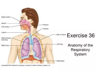
Lab Exercise 36 Anatomy of the Respiratory System Portland Community College BI 233
Terminology • Pulmonary Ventilation: aka breathing, is the movement of air into and out of the lungs • External Respiration: The gas exchange between the blood and alveoli • Internal Respiration: Exchange of gases between systemic blood and tissue cells • Cellular Respiration: Happens in Mitochondria Metabolic reactions that consume O2 and release CO2 during ATP production
Upper & Lower Respiratory System • Upper Respiratory System • Nose • Pharynx • Lower Respiratory System • Larynx • Trachea • Bronchi • Lungs
Nasal Cavity • The nasal epithelium covering the conchae serves to cleanse, warm and humidify the air • Nasal conchae increase the surface areas for the mucus epithelium • The olfactory epithelium in the upper medial part of the nasal cavity is involved in the sense of smell. • The nasal cavity serves as a resonating chamber as well as an avenue for escaping air.
Nasal Turbinates or Conchae • Ciliated pseudostratified columnar epithelium with goblet cells pushes trapped dust toward the back of the throat to be swallowed. .
Pharynx • Connects the nasal and oral cavities to the larynx and esophagus • Anatomically divided into 3 sections: • Nasopharynx • Oropharynx • Laryngopharynx
Tonsils Pharyngeal tonsils • Tonsils: lymphoid tissue. Palatine tonsils
Larynx: aka Voice Box • Made of 9 pieces of cartilage, the most important are: • Thyroid cartilage (Adam’s Apple) • Thyrohyoid membrane • Cricoid Cartilage • Cricothroid ligament • Epiglottis • Arytenoid Cartilage
Inside the Larynx • Vestibular Folds: folds of mucous membranes • Upper folds: false vocal cords • Lower folds: true vocal cords • These attach to the arytenoid cartilages by the vocal ligaments • Glottis: The vocal cords and the space between the folds. • Rima glottis: the space between the vocal folds
Trachea: aka Windpipe • Flexible and mobile tube extending from the larynx into the mediastinum • Composed of three layers • Mucosa: made up of goblet cells and ciliated pseudostratified columnar epithelium • Submucosa: connective tissue deep to the mucosa • Adventitia: outermost layer, has C-shaped rings of hyaline cartilage
Airways Larynx Trachea Right Mainstem Bronchi Left Mainstem Bronchi Secondary Bronchi Carina Secondary Bronchi
Branching of Bronchial Tree Trachea Primary Bronchi Secondary Bronchi Tertiary Bronchi Bronchioles Terminal/Respiratory Bronchioles
Bronchi • The carina of the last tracheal cartilage marks the end of the trachea and the beginning of the right and left bronchi • Left main stem bronchus • Right main stem bronchus • Bronchi subdivide into secondary bronchi, each supplying a lobe of the lungs
Bronchi Bronchioles • Tissue walls of bronchi mimic that of the trachea • As conducting tubes become smaller, structural changes occur and eventually they become bronchioles • Cartilage support structures change • Bronchioles differ from bronchi in that they lack cartilage • Epithelium types change • Amount of smooth muscle increases
Bronchioles Respiratory Bronchioles • Respiratory Bronchioles : Continued branching leads to the area where gas exchange occurs by simple diffusion
Bronchiole Histology Simple columnar epithelium Notice the lack of cartilage
Alveolar sacs Alveoli • Alveolar sacs look like clusters of grapes • The “individual grapes” are Alveoli
Type II Pneumocytes are cuboidal and produce surfactant Type 1 Pneumocytes are flattened for gas exchange
Respiratory Membrane • The area where gas exchange between air and blood occurs • It is the fused alveolar and capillary walls (3 layers) • Alveolar epithelium • Fused basal laminae • Capillary epithelium
Pleura • Pleura is the double-layered sac of serous membrane • Parietal Pleura is the outer layer and is attached to the thoracic walls • Visceral Pleura is the inner layer covering the lung tissue • The layers are only touching, they are not fused together • The potential space is called the pleural cavity • There is serous fluid between the layers which allows them to slide against each other during breathing
Lungs • Apex: the part under the clavicle • Base: the part touching the diaphragm • Costal Surface: the part touching the ribs • Hilus: indentation containing pulmonary and systemic blood vessels • Left Lung has 2 lobes and a cardiac notch • Left upper lobe • Left lower lobe • Right Lung has 3 lobes • Right upper lobe, middle lobe, lower lobe
Lungs LUL RUL Oblique fissure Horizontal fissure Cardiac notch Oblique fissure LLL RML RLL
Lab Exercise 37A Respiratory System Physiology Buffers
Inspiration/Expiration • Inspiration: Increase in thoracic cavity size • Inspiratory muscles • External intercostals (lift the rib cage) • Diaphragm (Becomes flat) • Expiration :Decrease in thoracic cavity size • Expiratory muscles • For the most part it is just the relaxation of the inspiratory muscles (passive process) • Internal intercostals & abdominal muscles used only for forced expiration
Respiratory Sounds • Bronchial sounds: Air in large passageways • Vesicular breathing sounds: air filling the alveolar sacs • Auscultation: • Throat • Intercostal spaces • Triangle of auscultation • Under the clavicle
Conducting Zone • All respiratory passageways that are not involved in gas exchange • All are mucous lined • From the nasal cavity to the terminal bronchioles • Aka: Anatomical dead space
Respiratory Zone • Thin walled simple squamous epithelium allows gas exchange with blood • Respiratory Zone Structures • Respiratory bronchioles • Alveolar ducts • Alveolar sacs • Alveoli
Spirometry • Spirometry is the classic pulmonary function test • Measures the volume of air inspired or expired as a function of time. • It can measure tidal volume, and vital capacity. • Spirometry may also be used to measure forced expiration rates and volumes and to compute FEV1/FVC ratios
Spirometry • Spirometry cannot access information about absolute lung volumes • It cannot measure the amount of air in the lung but only the amount entering or leaving.