Download
1 / 72
720 likes | 784 Views
The Immune System (rev 11/11). Defense mechanisms keep us healthy Physical and chemical barriers i.e. skin, stomach acids Prevent many harmful substances from entering the body and kill many that successfully entered

E N D
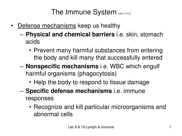
The Immune System (rev 11/11) • Defense mechanisms keep us healthy • Physical and chemical barriers i.e. skin, stomach acids • Prevent many harmful substances from entering the body and kill many that successfully entered • Nonspecific mechanisms i.e. WBC which engulf harmful organisms (phagocytosis) • Help the body to respond to tissue damage • Specific defense mechanisms i.e. immune responses • Recognize and kill particular microorganisms and abnormal cells Lec 9 & 10-Lymph & Immune
All three mechanisms work simultaneously to protect us. • A wide variety of cells, proteins, chemicals and organs are involved including the lymphatic system, the immune system, and the circulatory system. • Some microorganisms cause cell death and disease. These microbes are called pathogens. • These invade or poison living cells. • Release enzymes or toxins that damage cells, cause cell rupture, or invade cells and use the raw materials within the cell to duplicate themselves and starve or kill the healthy cell. Lec 9 & 10-Lymph & Immune
Eukaryotes (all organisms except bacteria) • Defining feature of eukaryotic cells is the membrane bound nucleus. • Nucleus contains chromosomes and associated proteins. • Have well organized internal structures called organelles. Lec 9 & 10-Lymph & Immune
Prokaryotes (bacteria) • Single celled • Do not have a nucleus or membrane bound organelles • DNA is contained in 1 chromosome • Outer surface is covered by a rigid cell wall that surrounds the plasma membrane and gives bacteria their shape (spheres, rods, spiral) • Use of variety of resources to get materials for energy, growth and reproduction Lec 9 & 10-Lymph & Immune
Prokaryotes: Viruses • Extremely small infectious agents • Structurally, consists of only a small piece of RNA or DNA, which contains the virus’ genetic material, surrounded by a protein coat • Has no organelles of its own so it can’t grow or reproduce on its own; forces the living cell to make more copies of the virus. • Viruses can only reproduce when they make contact with a living cell. They take over the living cell and use the cell’s organelles to reproduce more virus cells. Lec 9 & 10-Lymph & Immune
Modes of entry into living cells: • Taken into the cell cytoplasm by endocytosis. Once inside, the protein coat dissolves and the viral genetic material is released and incorporated into the cell’s genetic material • Merge their outer coat with the cell membrane and release the genetic contents into the cell’s cytoplasm • Attach to the outer surface of the cell membrane and inject their genetic material into the cell • Once inside the cell, the virus genetic material causes the cell to begin producing copies of the virus Lec 9 & 10-Lymph & Immune
Prions • Are infectious proteins that cause normal brain cell proteins to misfold and then build more prions. • The prions accumulate in brain cells until the cells malfunction or stop working completely. The infected cells die and burst, releasing prions to infect other brain cells. • Gradual build up of prions causes debilitating neurological symptoms and progressive degeneration. • No known cure available; prions resist cooking, freezing, drying • Disease is called Mad Cow disease in animals or Creutzfeldt-Jakob disease in humans. Both are fatal neurological disorders. Lec 9 & 10-Lymph & Immune
Determination of Health Risk Factors Which Determine the Danger from a Pathogen • Transmissibility: how easily it is passed from person to person • Mode of transmission: how it is transmitted • respiratory, fecal, oral, body fluids • Virulence: how damaging the resulting disease is Lec 9 & 10-Lymph & Immune
Lymphatic System The lymphatic system consists of : • Lymphatic vessels • Lymphoid tissues and organs throughout the body • Lymphatic vessels transport back to the blood any fluids which have escaped from the circulatory system. • Once interstitial fluid enters a lymphatic vessel, its name changes to lymph • Lymphatic tissues contain phagocytic cells and lymphocytes which play essential roles in the body’s defense mechanism and its resistance to diseases. • Lymph nodes-clean lymph as it passes through them Lec 9 & 10-Lymph & Immune
Lymphatic System Functions: • Protection against disease and injury • Filtration of foreign material to defend against infection and injury • Maintenance of blood volume in cardiovascular system • Capillaries are not watertight and some fluid leaks out into the tissues • Removes excess tissue fluid & returns it to the circulatory system • Transport of fats and fat-soluble material absorbed from the digestive system • Lymph activates the immune system. • Lymphocytes monitor the lymphatic stream for the presence of antigens and mount an attack against them. Lec 9 & 10-Lymph & Immune
Lymph: WBCs, fat cells, and protein-containing fluid transported by lymphatic vessels; Lymphatic vessels have: • Walls consisting of 3 thin layers • One way valves to prevent backflow of lymph (lymph flows only toward the heart) • Skeletal muscle contraction and pressure changes in the chest during breathing keep the lymph flowing Lec 9 & 10-Lymph & Immune
Lymph capillaries have wide spaces between its cells. This allows them to take up substances, including proteins, bacteria and cancer cells, that are too large to enter a blood capillary. These substances then use the lymphatic vessels to travel throughout the body. • Lymphatic capillaries are typically located throughout the blood capillary beds so they can pick up the fluid that has leaked out of the circulatory system into the tissue spaces. Lec 9 & 10-Lymph & Immune
As blood circulates through the body, nutrients, wastes and gasses are exchanged between the blood and the interstitial fluid. Fluid which remains in the tissue spaces becomes part of the interstitial fluid (approximately 3 liters). This leaked fluid and any plasma proteins that escape from the blood stream must be carried back to the blood. • As the fluid pressure in the tissue spaces increases, little flaps in the lymphatic capillaries are forced open, the excess fluid enters the lymphatic capillaries and will later be returned into the circulatory system to ensure there is sufficient blood volume for it to function properly. Lec 9 & 10-Lymph & Immune
Located at intervals along the lymphatic vessels are organs called lymph nodes. • Lymph nodes are the principal lymphatic organ in the body. • They filter and remove microorganisms, cellular debris, and abnormal cells from the lymph before returning the cleansed fluid to the cardiovascular system which prevents this from being delivered into the blood and spreading to other parts of the body • Lymph nodes cluster along the lymphatic vessels and most are buried in connective tissue so we can’t see them. • Large clusters of nodes occur near body surfaces in the inguinal (groin), axillary and cervical regions. Lec 9 & 10-Lymph & Immune
Lymph nodes have 2 basic functions: • act as lymph filters • they help activate the immune system. • Lymphocytes monitor the lymphatic stream for the presence of antigens and mount an attack against them. • Lymphatic capillaries merge to form small vessels larger and larger vessels trunks then create 2 major lymphatic ducts • the right lymphatic duct and the thoracic duct (right lymphatic duct drains the right arm and the right side of the head and chest; the thoracic duct is much larger and drains lymph from the rest of the body) • The right lymphactic duct drains into the right the subclavian vein and the thoracic duct into the left subclavian vein and return lymph to the cardiovascular system. Lec 9 & 10-Lymph & Immune
Lymphatic capillaries merge to form small vessels larger and larger vessels trunks then create 2 major lymphatic ducts • the right lymphatic duct and the thoracic duct (right lymphatic duct drains the right arm and the right side of the head and chest and drains into the right subclavian vein; -- the thoracic duct is much larger and drains lymph from the rest of the body and drains into the left subclavian vein ) Lec 9 & 10-Lymph & Immune
Other important lymphatic system structures: • Spleen, thymus gland, tonsils and adenoids Spleen---Largest lymphatic organ • Is a site for production of lymphocytes, usually in response to invading pathogens, and RBC • Has red pulp and white pulp tissue. • White pulp is lymphatic tissue • Red pulp is filled with blood as well as lymphocytes and macrophages (type of phagocyte) • Contains macrophages that • filter blood and trap bloodborne antigens; • scavenge and break down microorganisms, foreign matter, and old or damaged RBC and platelets • Saves hemoglobin (iron) from the RBC Lec 9 & 10-Lymph & Immune
Cleans and stores blood; • this blood is used in case of severe blood loss (hemorrhage) or a fall in blood pressure or whenever extra oxygen carrying capacity is needed. • Immune surveillance and response • Helps fight infection Lec 9 & 10-Lymph & Immune
Some Diseases Which Cause Spleen to Enlarge: Tuberculosis and leukemia • Frequently swollen spleen is felt as a lump in the upper left abdomen • A strong blow to the abdomen can rupture the spleen and cause severe internal bleeding. • May need a splenectomy to prevent hemorrhage • We can live without a spleen because its functions are shared by the • Lymph glands, liver, red bone marrow • But, people are more vulnerable to infection if they have had a splenectomy Lec 9 & 10-Lymph & Immune
If the spleen is removed, the lymph glands, liver and bone marrow take over most of its functions. • In children under 12, the spleen can regenerate if a small part of it is left in the body • Main distinction between spleen and lymph nodes is which fluid they clean • Spleen cleans the blood • Lymph nodes clean the lymph • Together they keep the circulating body fluids clean of damaged cells and microorganisms. Lec 9 & 10-Lymph & Immune
Thymus • Gland contains lymphocytes and epithelial cells • Secretes 2 hormones, thymosin and thymopoietin, that stimulate T lymphocytes (T cells) to mature and work against specific pathogens which invade the body • Gland is largest and most active during childhood and begins to shrink after adolescence since defense mechanisms are usually well established by then. Lec 9 & 10-Lymph & Immune
Tonsils • Lymphocytes in the tonsils gather and filter out many of the microorganisms that enter the throat in food or air • Tonsillitis and tonsillectomy: • Tonsils in the back of the throat are the largest and most often infected • If infection is serious, tonsillectomy may be needed Lec 9 & 10-Lymph & Immune
Adenoids • Also known as pharyngeal tonsils; are at the back of the nasal passages • Tend to enlarge during early childhood, begin to shrink after 5 years of age, and disappear by puberty • Sometimes they keep enlarging and block airflow from the nose to the throat • Causes mouth breathing, a nasal voice, and snoring • Remove them surgicallyadenoidectomy Lec 9 & 10-Lymph & Immune
Lines of Defense The lymphatic system works with other body systems to protect us against pathogens and cellular changes Remember the defense mechanisms which we spoke about earlier: • Physical and chemical barriers • Non-specific defense mechanisms • Specific defense mechanisms Lec 9 & 10-Lymph & Immune
Non-Specific Defense Systems First Line of Defense • Physical and chemical barriers • Physical barriers: prevent pathogens from entering the body • Chemical defenses produce substances that slow the growth or kill pathogens • The intact skin and mucous membranes are our most important barrier and first line of defense. • Structure: dead layer, inhospitable to microorganisms • Constant replacement and repair: many adhering microorganisms removed • Acidic pH (5–6): too acidic for many microorganisms; this inhibits their growth • Sweat glands produce antibiotic fluid (dermicidin) Lec 9 & 10-Lymph & Immune
Other: tears, saliva (saliva and tears secrete lysozyme, an enzyme which kills bacteria), earwax, digestive acids, hair, mucus (mucus is sticky and traps pathogens), vomiting, urination, defecation, resident bacteria (normal flora), mucous membranes (line all body cavities open to the exterior) • Most “successful” pathogens enter the body where we don’t have skin and they can take advantage of moist surfaces in direct contact with living cells. Lec 9 & 10-Lymph & Immune
Nonspecific Defenses: Second Line Antimicrobial proteins, phagocytes and other cells inhibit the spread of invaders throughout the body. • These defenses don’t target specific pathogens; they occur in response to any body threat • They work to actively seek out pathogens which have penetrated our physical and chemical barriers and are killing or damaging cells and then attack the invaders • Respond to tissue damage by removing debris Lec 9 & 10-Lymph & Immune
Nonspecific Defenses Phagocytosis: by neutrophils, macrophages (chief phagocyte), and eosinophils • Neutrophils (most abundant WBC) are the first WBC at the infection site. • Antimicrobial chemicals (defensins) are produced and pierce the pathogen’s cell membrane • They engulf, digest and destroy bacteria in the blood and tissue fluids • Remember that some WBC can filter through the walls of blood vessels into tissue spaces (they are attracted by substances released by injured cells at the infection site) Lec 9 & 10-Lymph & Immune
Monocytes leave the blood vessels, enter the tissue fluids, and develop into macrophages which engulf and digest large numbers of pathogens and anything foreign to the body and help to activate T cells. • Macrophages also cleanup by scavenging old blood cells, dead tissue fragments and other cellular debris. • They also release chemicals that stimulate the production of more WBC. • Eosinophils surround large invaders and bombard them with digestive enzymes as well as surround and digest some foreign proteins. Lec 9 & 10-Lymph & Immune
When the body is actively fighting an infection, many WBC die. • We also have tissue fluid, dead phagocytes, dead pathogens and cellular debris at the infection site. • These produce pus. • (neutrophils destroy themselves in the phagocytic process; macrophages don’t) • If the pus becomes trapped and can’t drain, the body forms an abscess. • It walls off the pus by forming a connective tissue “jail cell” for the pus. Lec 9 & 10-Lymph & Immune
Inflammatory response or Inflammation: • Triggered by any type of tissue injury • Ultimate goal of inflammation is to clear the injured area of pathogens, dead tissue cells, and any other debris to that the tissue can be repaired • Visible Signs: redness, warmth, swelling, pain, and sometimes, impairment of function so we rest the injured part • Tissue damage leads to release of chemicals from damaged cells. The chemicals stimulate mast cells, connective tissue cells specialized to release histamine. Histamine causes blood vessels to dilate so more blood flows into the area which causes the redness, warmth/heat, swelling. Lec 9 & 10-Lymph & Immune
Local capillaries also release fluid containing clotting factors and antibodies. This fluid goes into the tissue spaces, causing swelling and then pain from pressure on local nerve endings. • Swelling helps to dilute harmful substances and brings complement proteins and including clotting factors. • Clotting factors form a mesh foundation for permanent repair which helps to isolate the injured area and prevent the spread of bacteria to neighboring tissues. Lec 9 & 10-Lymph & Immune
Complement proteins • mark bacteria; • phagocytic cells arrive and remove invading microorganisms, try to prevent damage from spreading, • dispose of cellular debris, and • begin the tissue repair process. Lec 9 & 10-Lymph & Immune
Complement system • Group of plasma proteins that circulate, in an inactive state, in the blood and assist other defense mechanisms; they enhance or “complement” the effectiveness of the 1st and 3rd lines of defense • When presence of an infection activates a complement protein, the protein activates another and each protein keeps activating others. • Some complement protein cells join to form large protein complexes that create holes in (lyse) bacterial cell walls. Fluids and salts enter through the holes and the bacterium swells and bursts. (Normal cells have proteins to inactivate the complement.) • Others bind to bacterial cell membranes and mark them for destruction by phagocytes. • Others stimulate mast cells to release histamine or help get additional phagocytes to the infection site. Lec 9 & 10-Lymph & Immune
Second Line of Defense Natural killer (NK) cells: lymphocytes which destroy tumor cells and virus infected cells • They police the body via the blood and lymph andidentify invading cells by certain changes that take place in the plasma membranes (the lack of “self” cell surface receptors). • NK cells are non-specific killers (they do NOT target specific enemies). • NK cells are not phagocytes. They release chemicals that break down their targets’ cell membranes. • After an attack from NK cells, the “bad” cell membranes develop holes and the nucleus disintegrates. • NK cells also secrete substances that help the inflammatory response. Lec 9 & 10-Lymph & Immune
Interferons: interfere with virus spread • When a cell becomes virus infected, it will ultimately succumb to the virus, but it will try to help protect other normal cells by releasing a group of proteins called interferons. Antimicrobial proteins help other defenses by attacking the microorganism directly or by hindering their ability to reproduce. • Viruses lack the cellular machinery to make ATP or proteins. In order to survive, the viruses alter the host’s cell’s “machinery” so it will make more viruses. • Interferons diffuse to healthy cells, bind to their cell membranes, and stimulate the still healthy cell to produce proteins that interfere with the virus from making the proteins it needs to survive. This makes it harder for the virus to establish itself. • Interferons also activate microphages and mobilize natural killer cells. Lec 9 & 10-Lymph & Immune
Fever: increases host cell defenses and metabolic activity • When macrophages attack any foreign substance, they release pyrogens into the bloodstream and cause a fever • The higher temperature helps our body fight infection as well as makes the virus uncomfortable due to the heat. • Fever increases the metabolic rate and thus speeds up the defense mechanisms and tissue repair processes • An extremely high fever can be dangerous though because it can affect the shape of the chemical bonds that give proteins their shape and allow them to function. • (Inflammation is a localized response to infection) • Fever is a systemic response to invading microorganisms Lec 9 & 10-Lymph & Immune
Third Line: Specific Defense Mechanisms • Immune response—targets specific enemies, however it takes more time to mobilize than the non-specific defenses • Has 3 important characteristics: • Recognizes and targets specific pathogens or foreign substances • Has a “memory”—the ability to store information from past exposures so it can respond more quickly the next time the pathogen attacks • Protects the entire body; the resulting immunity is not limited to the infection site • Key to this response is the body’s ability to distinguish between its own cells and those of foreign invaders. • Defends the body by directly attacking cells as well as indirectly by releasing mobilizing chemicals and protective antibodies Lec 9 & 10-Lymph & Immune
Two separate but overlapping branches of this defense system 1. Humoral immunity: provided by antibodies present in the body’s “humors” or fluids—blood, lymph --antibodies circulate in the blood and lymph and bind to bacteria, bacterial toxins, and free viruses. The antibodies temporarily inactivate these microorganisms and mark them for destruction by phagocytes or complement. 2. Cell mediated immunity: lymphocytes defend the body by attacking the infected cells. Lymphocytes act directly to kill the foreign cells or indirectly by releasing chemical mediators that help the inflammatory response or activate other lymphocytes or macrophages Lec 9 & 10-Lymph & Immune
Antigens (antibody generating) • Any substance that mobilizes the immune system and causes an immune response. • Each antigen has a unique shape that allows the immune system to recognize it. • Antigens are usually a large protein or polysaccharide molecule • The immune system responds by making antibodies to attack and kill the antigen. Lec 9 & 10-Lymph & Immune
Human cells also have surface proteins which our immune system uses to recognize that the cells belong to you. These self-markers or self-antigens are called major histocompatibility complex (MHC) proteins. • They signal the immune system to not react to your own cells. • Some small molecules don’t cause an immune response, however if they link up with the body’s proteins, the immune system may recognize them as foreign and start an attack, causing hypersensitivities i.e. allergies, medicine reactions, poison ivy reaction, etc. • Transplant reactions • Abnormal and cancerous cells have MHC proteins that are not recognized as “self”. Lec 9 & 10-Lymph & Immune
Lymphocytesare the main fighter of the immune system and protect the body against antigens. • They come from the red bone marrow and mature into immunocompetent cells, either T cells (T lymphocytes) or B cells (B lymphocytes). They are named forwhere they mature— T cells mature in the thymus gland; B cells mature in bone marrow • Both recognize and target antigen bearing cells differently. Activated T cells manage the immune response and some of them directly attack and destroy infected cells . B cells produce plasma cells which secrete antibodies into the blood and other body fluids. Lec 9 & 10-Lymph & Immune
The immune system’s response to threats to the body depends on the ability of its cells to recognize antigens and bind to them and to communicate with one another so that the whole system mounts a response specific to those antigens. Lec 9 & 10-Lymph & Immune
B cells are responsible for antibody-mediated immunity. B cells produce antibodies (proteins that bind with and neutralize specific antigens.) They release antibodies into the lymph, blood, and tissue fluid so they can circulate throughout the body. Antibodies immobilize antigens until they can be destroyed by phagocytes or other means. In adults, B cells mature in bone marrow. When they mature, they develop surface receptors that allow them to recognize specific antigens. • They travel in the blood to the lymph nodes, spleen and tonsils and remain inactive until they meet a foreign cell with the specific antigen to which they respond. Lec 9 & 10-Lymph & Immune
The foreign cell activates the B cell to grow and multiply. The resulting group of identical cells is called a clone. • Clone cells are called plasma cells becausethey secrete their antibodies into the lymph fluid and into the blood plasma. • Antibodies are made and continue to circulate in our blood. • When the antibodies meets the correct antigen, they bind to them and create an antigen-antibody complex. • This complex marks the antigen and the cell carrying it for destruction. • Macrophages and activated B cells engulf foreign particles and digest them. Lec 9 & 10-Lymph & Immune
Some of the clone cells become memory cells. These cells remain inactive in our body until the same antigen reappears. • Memory cells store information about the pathogen; if we have a second exposure, the immune response is faster because the memory cells react immediately to the threat. Lec 9 & 10-Lymph & Immune
Antibodies: • Neutralize non-self antigens • Their “Y” shape allows them to “lock” into a cell and attack its contents. • Five classes of antibodies: IgG, IgM, IgA, IgD, IgE • Antibodies belong to the class of blood plasma proteins called gamma globulins However, they are referred to as immunoglobulins (Ig) because of the crucial role they play in immunity. Lec 9 & 10-Lymph & Immune
Immunoglobulins Antibodies are also called immunoglobulins; they are the gamma globulin part of blood proteins. Antibodies are proteins secreted by activated B cells or plasma cells in response to an antigen and are capable of binding specifically with the antigen. Five major classes: (MADGE is a mneumonic) • IgG-75% • This is the most abundant antibody. They are the only antibodies that cross the placenta (during pregnancy) and pass on the mother’s immunities to the fetus. • IgM-5-10% • These are the first to be released during immune responses. They cause cells to clot. ABO blood cell antibodies belong to this class. Lec 9 & 10-Lymph & Immune
IgA-15% • Play a major role in preventing pathogens from entering the body. • These enter areas of the body covered by mucous membranes where they neutralize pathogens. They are present in mother’s milk and are passed on to the infant during breast feeding. • IgD-<1% • Their function is unclear. • IgE-0.1% • These activate the inflammatory response by causing the release of histamine. They also cause allergic responses. • Antibodies are specialized to latch onto intact bacteria and foreign molecules in the extracellular environment (body secretions, tissue fluid), as well as circulating in blood and lymph. Lec 9 & 10-Lymph & Immune
T cells • Circulate continuously throughout the body • Are responsible for cell-mediated immunity which depends upon the actions of several types of T cells. • T cells don’t produce antibodies; the cells directly attack foreign cells that carry antigens or they release proteins that boost other aspects of the immune response. • T cells can identify and kill infected human cells before the cells have a chance to release new pathogens into the blood. Lec 9 & 10-Lymph & Immune