Download
1 / 35
390 likes | 780 Views
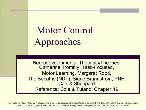
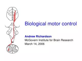
Motor control. Proprioception and movement. 2 /24. in general we are not aware of information coming from proprioceptors though they belong to somatosensory receptors

E N D
Proprioception and movement 2/24 • in general we are not aware of information coming from proprioceptors though they belong to somatosensory receptors • these receptors detect stretching of the muscles and tension in tendons and induce reflexes as well as provide information for the control of movement • receptors in joints detecting the angle of the joints also belong to this category • they also provide input for the control of movement • these facts justify treatment of these receptors in the framework motor control
Final common pathway 3/24 • somatic and visceralmotor systems represent the output of the CNS • in the somatic system the final common pathway is the motor neuron in the ventral horn of the spinal cord or in the motor nucleus of cranial nerves in the brainstem • in the visceral system the final common pathway means the motor neuron in the lateral horn of the spinal cord or in the vegetative nucleus of cranial nerves in the brainstem • final common pathway means (Sherington) that executive organs can be only accessed through these motor neurons, integration occurs at this or at a higher level • in the somatic system, fibers innervate targets (skeletal muscle) directly, in the visceral (smooth muscle and gland cells) through an intercalated neuron
Hierarchical organization 4/24 • areas where stimulation causes movements are classified as motor areas • these areas receive somatosensory input as well, thus they are sometimes referred to as somatomotor areas and systems • regulation is done on several levels – the higher the level the more complicated movements are controlled, though lower levels are influenced by descending effects as well • cortical areas are able to control motor neurons in the spinal cord and brainstem directly, but they also exert their effects on the spinal cord indirectly through the brain stem • in addition to these pathways, cerebellum and basal ganglia also participate in motor control by influencing brain stem and cortical areas • somatotopy is present at every level
Organization of motor system cortex basal ganglia thalamus cerebellum brain stem motor neuron sensory pathway brain stem and spinal cord 5/24
Muscle spindle 6/24 • muscle spindle is 4-10 mm long, it is made up by 6-8 modified muscle fibers, and is located in parallel with regular muscle fibers • muscle spindle is encased in a connective tissue capsule • within the muscle spindle, the middle part of intrafusal fibers is modified for receptor function, terminal parts are able to contract • usually there are 2 nuclear bag (one dynamic, one static)and 5-6 nuclear chain fibers (all static) in a spindle • primary terminals form spirals around the middle part of all three types of receptors - annulospiral terminal - Ia (Aα) afferent • secondary terminals (flower spray endings) target static nuclear bag and nuclear chain fibers - II (Aβ) afferent Berne and Levy, Mosby Year Book Inc, 1993, Fig. 12-2 Berne and Levy, Mosby Year Book Inc, 1993, Fig. 12-1
Operation of the muscle spindle • when extrafusal (regular) fibers contract, intrafusal fibers are shortened and relax • when extrafusal fibers are stretched, then intrafusal fibers are also stretched – elongation of receptor terminals increase the discharge rate • in case of continuous stretch polar regions of dynamic nuclear bag receptors lengthen due to their viscoelastic properties, thus excitation of the nerve terminal decreases • regular muscle fibers are innervated by A axons, intrafusal fibers by A axons – excitation is parallel, thus sensitivity of the receptor remains constant • previously a servo-mechanism was suggested 7/24
The tendon organ 8/24 • tendon organs are in series with muscles • they are 1 mm long structures surrounded by a capsule • collagen fibers of the tendon penetrate the muscle • perpendicularly Ib afferents, branches run between fibers or wrap around them • they are deformed with increasing tension – excitation • tendon organs inform about the contraction and passive extension of muscles, but they are more sensitive to the former • thus, they provide information about forces developing in the muscles
Spinal reflexes I. 9/24 • the lowest level in the hierarchy of the motor control is represented by the spinal reflexes • the myotatic (stretch) reflex is monosynaptic: afferents terminate on the motoneuron of the same muscle – proprius (Latin): one’s own • stretching of muscle spindle induces contraction of the same muscle • patella reflex, Achilles tendon reflex – mostly in extensors, though in flexors as well (biceps) • collateral of Ia afferent inhibits antagonist motoneuron through an inhibitory interneuron – reciprocal innervation is characteristic for the spinal cord – simultaneous contraction is organized always at a higher level • myotatic reflex can be dynamic or tonic, in the second type secondary terminals also participate Berne and Levy, Mosby Year Book Inc, 1993, Fig. 12-11
Spinal reflexes II. 10/24 • diagnostic value: motoneuron excitability – direct stimulation: H or Hoffman reflex • its function is to keep posture, thus it is stronger in extensors than in flexors, but sloth • divergence and convergence in respect of agonist muscles are both present – though the reflex remains segmental • muscle tone (resistance against passive moving) is due to this reflex: a subset of motor units are always slightly contracted • myotatic reflex, thus muscle tone is continuously modified by descending effects through setting the sensitivity of the motoneuron (e.g. REM) • collateral of Ia afferent terminates on neurons belonging to the column of Clarke: spinocerebellar tract
Spinal reflexes III. 11/24 • inverse myotatic reflex starts from tendon organs • incoming fibers target agonist motoneuron through inhibitory interneurons, antagonist through excitatory ones • main function is to protect muscle and tendon against overstretching • it also supplements myotatic reflex: when tension in tendon decreases – weaker inhibition – contraction • flexion withdrawal reflex starts from nociceptors (exteroceptive), its function is to remove extremity from the noxious stimulus source • it is polysynaptic, in addition to activate antagonist muscles, it also induces crossed extension reflex • the reflex is intersegmental, many muscle groups can participate Berne and Levy, Mosby Year Book Inc, 1993, Fig. 12-13 Berne and Levy, Mosby Year Book Inc, 1993, Fig. 12-12
Motor stereotypes 12/24 • spinal cord is able to organize simple motor stereotypes – these are intersegmental • scratching reflex involves rhythmically alternating movements, frequency is independent from the strength of the stimulus, it only increases duration • afterdischarge is characteristic (reverberating circuits) • stereotypes are generated by a central rhythm generator no feedback from proprioceptors is needed – based on mutual inhibition, adaptation and rebound • walking has similar central organization, but feedback from proprioceptors is needed and central descending effects influence frequency (walk, trot, canter, gallop)
Spinal cord organization I. 13/24 • location of motor neurons follows somatototopy • motor neurons of proximal and distal muscles are located medially and laterally, respectively • axial muscles in the midline of the trunk belong to the most medial motor neurons • these motor neurons receive input from interneurons on both sides – bilateral control – posture • motor neurons of extensors and flexors are located ventrally and dorsally, respectively • motor neurons controlling a given muscle are found in 1-4 neighboring segments – motor neuron pool • within the pool muscle fibers innervated by 1 motor neuron form the motor unit – 10 (eye), 100 (hand), 2000 (foot) fibers • all fibers in a unit are the same type
Types of muscle fibers 14/24 • tonic fibers • postural muscles in amphibians, reptiles and birds • muscle spindles and extraocular muscles in mammals • no AP, motor axon forms repeated synapses • slow shortening – effective isometric contraction • slow-twitch (type I) fibers • mammalian postural muscles • slow shortening, slow fatigue – high myoglobin content, large number of mitochondria, rich blood supply – red muscle • fast-twitch oxidative (type IIa) fibers • specialized for rapid, repetitive movements – flight muscles of migratory birds • many mitochondria, relatively resistant to fatigue • fast-twitch glycolytic (type IIb) fibers • very fast contraction, quick fatigue • few mitochondria, relies on glycolysis • breast muscles of domestic fowl – white muscle
Spinal cord organization II. 15/24 • reflexes activate only part of the motor units – fractionization principle • reflexes and voluntary movements can be graded – more and more units get involved – recruitment • activation follows size principle – first small units are activated – motor neurons are also small, EPSPs are more effective • the largest units contain fast-twitch glycolytic fibers (white muscle) – they are only activated when really necessary • in addition to recruitment, frequency can be also increased - during voluntary movements 8-25 Hz causing incomplete tetanus – motor units contract asynchronously
Inhibitory interneurons 16/24 • α-motor neurons are innervated by three types of inhibitory interneurons: Ia, Ib and Renshaw • Ia receives input from the muscle spindle in the antagonist muscle, but it is also activated by descending fibers targeting the antagonist motor neuron • it also receives inhibitory inputs – suspension of reciprocal innervation – „column” function • Ib receives input from the tendon organ, but it is also influenced by descending pathways, receptors in the skin and joints – these control the strength of contraction: touching, stroking • Renshaw receives input from collaterals of α-motor neurons – feedback inhibition • sensitivity is controlled by descending excitatory and inhibitory pathways
Brain stem reflexes and posture • lesions of the neuraxis change the tone of postural muscles – Sherington: tone in these muscles is caused by reflexes • tone is modified by descending effects: lifting one leg increases the tone of the others • transection between the n.ruber and the Deiters’ nucleus – decerebrate rigidity in tetrapods • it can be abolished by cutting the reflex arch • Deiters’ nucleus (tr. vestibulospinalis lat.) and pontine RF (tr. reticulospinalis med.) strongly enhances extensor tone • inhibitory effects: • cerebellum • in tetrapods tr. rubrospinalis from n. ruber • in primates it has only effect to the level of cervical segments, cortex is more important • medullary RF – tr. reticulospinalis lateralis 17/24
Voluntary movements I. 18/24 • the background for voluntary movements is provided by muscle tone control • appropriate rearrangement of muscle tone is needed to preserve posture during voluntary, reflex and stereotype movements • common characteristic of voluntary movements that they become automated through learning and exercise – learning to walk in babies sports, etc. • Fritsch and Hitzig 1870: cortical stimulation in dogs might lead to movements • information about the organization of cortical motor control was collected from five sources: • stimulation studies (Penfield human surgeries) • analysis of brain injuries • unit recording in monkeys • imaging, e.g. PET • anatomical studies
Voluntary movements II. 19/24 • based on these data we know what type of damages impair control of voluntary movements, and which areas are activated first – organization of motor control is less clear • primary motor area: Br.4 – gyrus precentralis • somatotopy is similar to the somatosensory area: leg medially, representation is proportional with the sophistication of movements • secondary motor cortex: Br.6 – in front of primary • it consists of two parts: supplementary motor area and premotor cortex • their role is in the preparation (premotor), and in the planning (supplementary) of movements: electrophysiological and blood flow changes before the movements and during contemplating of movements Blumenfeld, Sineauer Assoc. Inc., 2002, Fig. 2-13
Voluntary movements III. 20/24 • the corticospinal or pyramidal tract is the most important motor pathway • most of the fibers originate from layer V pyramidal neurons in Br.4 and 6, but from other areas as well – upper motor neurons • 90% of the axons cross over and run in the „pyramids” on the surface of medulla (name), then in the lateral corticospinal tract, 10% cross in the spinal cord (ant. tr.) before ending • direct effect on α-motor neurons (lower motor neurons), indirect effect through interneurons • motor cortices receive input from the VL (thalamus) and from the somatosensory cortex • VL transmits information from cerebellum and putamen, no direct projection • interaction goes both ways (see before) • movements can be elicited from other cortical areas as well, but with strong stimuli only
Cerebellum I. 21/24 • cerebellum coordinates motor activities, its injuries impair coordination and execution of voluntary movements • motor learning is also lost • cerebellum has more neurons than the other parts of the CNS • modular structure, János Szentágothai contributed heavily to its description • principal neurons are inhibitory Purkinje cells, projecting to deep cerebellar nuclei that in turn project to VL • various excitatory (e.g. granule) and inhibitory (e.g. Golgi) interneurons • input: climbing fiber (contralateral oliva inferior) and mossy fiber (cortex, spinal cord, brainstem) • multiple somatotopical representation in the cortex and deep cerebellar nuclei Berne and Levy, Mosby Year Book Inc, 1993, Fig. 14-9 Berne and Levy, Mosby Year Book Inc, 1993, Fig. 14-12
Cerebellum II. 22/24 • three parts (connections, evolution): • vestibulocerebellum (archeocerebellum) – oldest, caudal part (flocculus, nodulus) • direct input from semicircular canals, utriculus, sacculus • direct output to Deiters’ nucleus (it can be considered as a deep cerebellar nucleus) • balance and gait, coordination of eye movements, reflex movements of the head • spinocerebellum (paleocerebellum) – middle part of cerebellum (vermis, central, intermediary parts of the hemispheres) • input through the dorsal spinocerebellar tract from sensory afferents – information about the position of extremities and about changes that occurred (external feedback) • input through the ventral spinocerebellar tract about the activity of interneurons: it reflects descending commands (internal feedback) • the cerebellum monitors the execution of motor commands • cerebrocerebellum (neocerebellum) – lateral part of hemispheres • input from the cortex and n. ruber through the pons • output to n. dentatus, thalamus, cortex • planning, starting and stopping as well as learning of movements Berne and Levy, Mosby Year Book Inc, 1993, Fig. 14-11
Basal ganglia I. 23/24 • basal ganglia consist of: • neostriatum (n. caudatus + putamen) • pallidum or globus pallidus (neostriatum+pallidum= corpus striatum, putamen+pallidum= n.lentiformis) • substantia nigra (pars compacta and pars reticularis) • n. subthalamicus • main functions are in the control of movements and muscle tone – deducted from impairments caused by lost of different cell groups • Parkinson’s disease: muscle rigidity, tremor, slowing or loss of physical movements (see Awakenings) – caused by loss of dopaminergic cell in substantia nigra pars compacta – MPTP (methyl-phenyl-tetrahydropyridine) • Huntington’s chorea: abnormal, jerky, random movements (chorea: Greek word for dance) – cholinergic and GABAergic neurons in the neostriatum die – genetic background is known – prenatal diagnosis – ethical issues
Basal ganglia II. 24/24 • outdated conception: pyramidal – extrapyramidal pathways • extrapyramidal was supposed to originate from the neostriatum – incorrect, the term is rarely used now • neurons of the basal ganglia are not activated before cortical neurons – no role in initiating movements • they modify through multiple circuits involving thalamic VA (ventralis anterior), VL (ventralis lateralis) and CM (centre median) the functioning of the motor cortex • in Parkinson’s disease stimulation of the direct and inhibition of the indirect pathway decrease – less excitation on VA, VL • in Huntigton’s chorea the indirect pathway is affected more - VA, VL inhibition decreases Berne and Levy, Mosby Year Book Inc, 1993, Fig. 14-21
Muscle spindle Berne and Levy, Mosby Year Book Inc, 1993, Fig. 12-1
Afferents in the muscle spindle Berne and Levy, Mosby Year Book Inc, 1993, Fig. 12-2
Myotatic reflex Berne and Levy, Mosby Year Book Inc, 1993, Fig. 12-11
Inverse myotatic reflex Berne and Levy, Mosby Year Book Inc, 1993, Fig. 12-12
Flexion withdrawal reflex Berne and Levy, Mosby Year Book Inc, 1993, Fig. 12-13
Somatomotor cortex Blumenfeld, Sineauer Assoc. Inc., 2002, Fig. 2-13
Cerebellum Berne and Levy, Mosby Year Book Inc, 1993, Fig. 14-9
Divisions of the cerebellum Berne and Levy, Mosby Year Book Inc, 1993, Fig. 14-11
Somatotopy in the cerebellum Berne and Levy, Mosby Year Book Inc, 1993, Fig. 14-12
Direct and indirect pathways Berne and Levy, Mosby Year Book Inc, 1993, Fig. 14-21