Immuno-diagnostics
Immuno-diagnostics. Dr. Kalpita Mulye Department of Biotechnology and Microbiology B. N. Bandodkar College. Modern methods. Basis: Specificity of Ag –Ab interaction Affinity and avidity Reversibility Cross reactivity (?) Advantages Highly Sensitive Precise Rapid Reproducible


Immuno-diagnostics
E N D
Presentation Transcript
Immuno-diagnostics Dr. Kalpita Mulye Department of Biotechnology and Microbiology B. N. Bandodkar College
Modern methods • Basis: • Specificity of Ag –Ab interaction • Affinity and avidity • Reversibility • Cross reactivity (?) • Advantages • Highly Sensitive • Precise • Rapid • Reproducible • Automated
Assay (microgm Ab/ml) • Precipitation reaction in fluids 20–200 • Precipitation reactions in gels • Mancini radial immunodiffusion 10–50 • Ouchterlony double immunodiffusion 20–200 • Immunoelectrophoresis 20–200 • Rocket electrophoresis 2 • Agglutination reactions • Direct 0.3 • Passive agglutination 0.006–0.06 • Agglutination inhibition 0.006–0.06 • Radioimmunoassay 0.0006–0.006 • Enzyme-linked immunosorbent assay (ELISA) 0.0001–0.01 • ELISA using • chemiluminescence 0.0001–0.01† • Immunofluorescence 1.0 • Flow cytometry 0.06–0.006
ELISPOT A well is coated with antibody against the antigen of interest, a cytokine in this example, and then a suspension of a cell population thought to contain some members synthesizing and secreting the cytokine are layered onto the bottom of the well and incubated. Most of the cytokine molecules secreted by a particular cell react with nearby well-bound antibodies.
After the incubation, the well is washed and an enzyme-labeled anti-cytokine antibody is added. After washing away unbound antibody, a chromogenic substrate that forms an insoluble colored product is added. • The colored product (purple) precipitates and forms a spot only on the areas of the well where cytokine-secreting cells had been deposited. • By counting the number of colored spots, it is possible to determine how many cytokine-secreting cells were present in the added cell suspension
RIA • Principle of RIA involves competitive binding of radio-labeled antigen and unlabeled antigen to a high-affinity antibody. • Direct binding assay • Hormone level detection • Need elaborate standardization process with pure antigen or antibody
Flow Cytometry • the flow cytometer, which was designed to automate the analysis and separation of cells stained with fluorescent antibody. • uses a laser beam and light detector to count single intact cells in suspension • instrument counts each cell as it passes the laser beam and records the level of fluorescence the cell emits; an attached computer generates plots of the • number of cells as the ordinate and their fluorescence intensity
FACS Every time a cell passes the laser beam, light is deflected from the detector, and this interruption of the laser signal is recorded. Those cells having a fluorescently tagged antibody bound to their cell surface antigens are excited by the laser and emit light that is recorded by a second detector system located at a right angle to the laser beam. capable of sorting populations of cells into different containers according to their fluorescence profile.
Use of the instrument to determine which and how many members of a cell population bind fluorescently labeled antibodies is called analysis; • use of the instrument to place cells having different patterns of reactivity into different containers is called cell sorting.
Immuno-hematology • Determination of blood group • Major and minor match • Isoagglutinin titre • Erythroblastosis fetalis
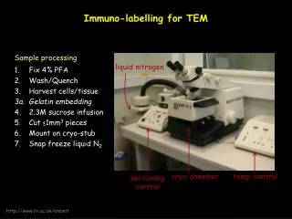
Immunoelectron microscopy • To detect intracellular location of structures or particular protein • High resolution • Specific antibodies are labeled with gold particles applied to ultra thin sections and observed in TEM • A number of electron-dense labels have been employed, including ferritin and colloidal gold. Because the electron-dense label absorbs electrons, it can be visualized with the electron microscope as small black dots.
Immuno fluorescence • Locate the target molecule in cells, tissues or biological fluids • Fluorescent antibodies: invaluable probes • First used for detecting plasma cells (Robin Coon) • Direct immuno fluorescence • Indirect immuno fluorescence
Advancements • Confocal fluorescent microscope • Time lapse video microscopy
ImmunohistochemistryIHC • Specific antibodies chemically coupled to an enzyme that converts the colorless substrate into colored reaction product in situ. • Tissue sectioning/ microtome • Fixation • Deparaffinisation • Use of polyclonal / monoclonal abs