Download
1 / 58
580 likes | 757 Views
2. Cells: The Living Units. C H A P T E R. Goals. By the end of this lecture you should be able to describe …. Similarities and differences between cells Why cells look and function differently The function of organelles in a typical eukaryotic cell How proteins are made

E N D
2 Cells: The Living Units C H A P T E R
Goals By the end of this lecture you should be able to describe …. • Similarities and differences between cells • Why cells look and function differently • The function of organelles in a typical eukaryotic cell • How proteins are made • How cells divide
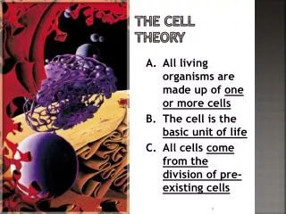
The cell theory: • Smallest living units in the body. • Perform all functions necessary to sustain life. • Obtain nutrients from surrounding body fluids • Disposes of its wastes and maintains its shape and integrity • Produced by the division of preexisting cells – they can replicate themselves
Introduction to Cells • Organelles – “little organs” – carry on essential functions of cells • Enzymes – direct chemical reactions in cells • Metabolism – the sum of all chemical reactions in the cell
Cells have three main components • Plasma membrane • Cytoplasm • Nucleus
The Plasma Membrane • Fluid mosaic model (lipid bilayer) • Types of membrane proteins • Integral proteins – firmly imbedded in, or attached to lipid bilayer • Peripheral proteins – attach to membrane surface
The Plasma Membrane Polar head of phospholipid molecule Extracellular fluid (watery environment) Glycolipid Cholesterol Nonpolar tail of phospholipid molecule Glycoprotein Carbohydrate of glycocalyx Bimolecular lipid layer containing proteins Outward- facing layer of phospholipids Inward-facing layer of phospholipids Integral proteins Cytoplasm (watery environment) Filament of cytoskeleton Peripheral proteins Figure 2.2
Functions of the Plasma Membrane • Physical isolation • Regulation of exchange with the environment • Sensitivity • Structural support
The Plasma Membrane • Determines which substances enter or leave the cell • Membrane is selectively permeable • Diffusion – molecules move from a region where they are more concentrated to an area where they are less concentrated • Osmosis – the diffusion of water across a membrane
Endocytosis • Mechanism by which particles enter cells • Phagocytosis – “cell eating” • Pinocytosis – “cell drinking” • Receptor-mediated endocytosis
Three Types of Endocytosis (b) Pinocytosis The cell “gulps” drops of extracellular fluid containing solutes into tiny vesicles. No receptors are used, so the process is nonspecific. Most vesicles are protein- coated. (a) Phagocytosis The cell engulfs a large particle by forming pro- jecting pseudopods (”false feet”) around it and en- closing it within a membrane sac called a phagosome. The phagosome then combines with a lysosome, and its contents are digested. Vesicle may or may not be protein-coated but has receptors capable of binding to microorganisms or solid particles. Vesicle Phagosome (c) Receptor-mediated endocytosis Extracellular substances bind to specific receptor proteins in regions of protein-coated pits, enabling the cell to ingest and concentrate specific substances in protein-coated vesicles. The ingested substance may simply be released inside the cell, or combined with a lysosome to digest contents. Receptors are recycled to the plasma membrane in vesicles. Vesicle Receptor recycled to plasma membrane Figure 2.4
Exocytosis Mechanism that moves substances out of the cell Substance is enclosed in a vesicle The vesicle migrates to the plasma membrane Proteins from the vesicles (v-SNAREs) bind with membrane proteins (t-SNAREs) The lipid layers from both membranes bind, and the vesicle releases its contents to the outside of the cell
Exocytosis (a) The process of exocytosis Plasma membrane SNARE (t-SNARE) Extracellular fluid 2 There, proteins at the vesicle surface (v-SNAREs) bind with t-SNAREs (plasma membrane proteins). Fused v- and t-SNAREs 1 The membrane- bound vesicle migrates to the plasma membrane. Secretory vesicle Vesicle SNARE (v-SNARE) Molecule to be secreted Cytoplasm Fusion pore formed 4 Vesicle contents are released to the cell exterior. 3 The vesicle and plasma membrane fuse and a pore opens up. Figure 2.5
The Cytoplasm • Cytoplasm – lies internal to plasma membrane • Consists of cytosol, organelles, and inclusions • Cytosol (cytoplasmic matrix) • Jelly-like fluid in which other cellular elements are suspended • Consists of water, ions, and enzymes
Cytoplasmic Organelles • Ribosomes – constructed of proteins and ribosomal RNA • Site of protein synthesis
Cytoplasmic Organelles • Endoplasmic reticulum – “network within the cytoplasm” • Rough ER – ribosomes stud the external surfaces • Smooth ER – consists of tubules in a branching network • No ribosomes are attached; therefore no protein synthesis
Cytoplasmic Organelles • Golgi apparatus – a stack of three to ten disk-shaped envelopes • Sorts products of rough ER and sends them to proper destination
Role of the Golgi Apparatus in Packaging Products of Rough ER
Mitochondria • Mitochondria – generate most of the cell’s energy; most complex organelle
Cytoplasmic Organelles • Lysosomes – membrane-walled sacs containing digestive enzymes • Digest unwanted substances • Peroxisomes – membrane-walled sacs of oxidase enzymes • Enzymes neutralize free radicals and break down poisons • Break down long chains of fatty acids • Are numerous in the liver and kidneys
Cytoplasmic Organelles • Cytoskeleton – “cell skeleton” – an elaborate network of rods • Contains three types of rods • Microtubules – cylindrical structures made of proteins • Microfilaments – filaments of contractile protein actin • Intermediate filaments – protein fibers
Centrosomes and centrioles • Centrosome – a spherical structure in the cytoplasm • Composed of centrosome matrix and centrioles • Centrioles – paired cylindrical bodies • Consists of 27 short microtubules • Act in forming cilia
Cytoplasmic Inclusions • Temporary structures – not present in all cell types • May consist of pigments, crystals of protein, and food stores • Lipid droplets – found in liver cell and fat cells • Glycosomes – store sugar in the form of glycogen
The Nucleus • The nucleus – “central core” or “kernel” – control center of cell • DNA directs the cell’s activities • Nucleus is approximate 5µm in diameter
The Nucleus Surface of nuclear envelope. Fracture line of outer membrane Nuclear pores Nuclear envelope Nucleus Chromatin (condensed) Nucleolus Nuclear lamina. The netlike lamina composed of intermediate filaments formed by lamins lines the inner surface of the nuclear envelope. Nuclear pore complexes. Each pore is ringed by protein particles Cisternae of rough ER (b) (a) Figure 2.13
The Nucleus • Nuclear envelope – two parallel membranes separated by fluid-filled space • Chromatin – composed of DNA and histone proteins • Condensed chromatin – contains tightly coiled strands of DNA • Extended chromatin – contains uncoiled strands of DNA • DNA's genetic code is copied onto mRNA (transcription)
The Nucleus • Chromosomes – highest level of organization of chromatin • Contains a long molecule of DNA
The Nucleus • Nucleolus – “little nucleus” – in the center of the nucleus • Contains parts of several chromosomes • Site of ribosome subunit manufacture
1 DNA double helix (2-nm diameter) Histones 2 Chromatin (“beads on a string”) structure with nucleosomes Linker DNA Nucleosome (10-nm diameter; eight histone proteins wrapped by two winds of the DNA double helix) (a) 3 Tight helical fiber (30-nm diameter) 4 Looped domain structure (300-nm diameter) 5 Chromatid (700-nm diameter) Metaphase chromosome (at midpoint of cell division) (b) Figure 2.15
The Cell Life Cycle • Is the series of changes a cell goes through • Interphase • G1 phase – growth 1 phase – the first part of interphase • Centrioles begin to replicate near the end of G1
The Cell Life Cycle • S (synthetic) phase – DNA replicates itself • Ensures that daughter cells receive identical copies of the genetic material • G2 phase – growth 2 phase– centrioles finish copying themselves • During S (synthetic) and G2 phases – cell carries on normal activities
The Cell Life Cycle G1 checkpoint (restriction point) Interphase S Growth and DNA synthesis G2 Growth and final preparations for division G1 Growth M Mitosis Prophase Metaphase Cytokinesis Telophase Anaphase Mitotic phase (M) G2 checkpoint Figure 2.16
The Cell Life Cycle • Cell division • M (mitotic) phase – cells divide during this stage • Follows interphase
The Cell Life Cycle • Cell division involves: • Mitosis – division of the nucleus during cell division • Chromosomes are distributed to the two daughter nuclei • Cytokinesis – division of the cytoplasm • Occurs after the nucleus divides
The Stages of Mitosis • Prophase – the first and longest stage of mitosis • Early prophase – chromatin threads condense into chromosomes • Chromosomes are made up of two threads called chromatids • Chromatids are held together by the centromere • Centriole pairs separate from one another • The mitotic spindle forms
The Stages of Mitosis • Prophase (continued) • Late prophase – centrioles continue moving away from each other • Nuclear membrane fragments
The Stages of Mitosis • Metaphase – the second stage of mitosis • Chromosomes cluster at the middle of the cell • Centromeres are aligned along the equator • Anaphase – the third and shortest stage of mitosis • Centromeres of chromosomes split
The Stages of Mitosis • Telophase – begins as chromosomal movement stops • Chromosomes at opposite poles of the cell uncoil • Resume their thread-like extended-chromatin form • A new nuclear membrane forms • Cytokinesis – completes the division of the cell into two daughter cells
Early Prophase and Late Prophase Late Prophase Interphase Early Prophase Early mitotic spindle Polar microtubule Centrosomes (each has 2 centrioles) Spindle pole Plasma membrane Fragments of nuclear envelope Aster Nucleolus Chromatin Centromere Chromosome consisting of two sister chromatids Kinetochore Kinetochore microtubule Nuclear envelope Figure 2.17 (1 of 2)
Metaphase and Anaphase Metaphase Anaphase Telophase and Cytokinesis Contractile ring at cleavage furrow Nucleolus forming Nuclear envelope forming Spindle Metaphase plate Daughter chromosomes Figure 2.17 (2 of 2)
Cellular Diversity • Specialized functions of cells relates to: • Shape of cell • Arrangement of organelles
Cellular Diversity • Cells that connect body parts or cover organs • Fibroblast – makes and secretes protein component of fibers • Erythrocyte – concave shape provides surface area for uptake of the respiratory gases • Epithelial cell – hexagonal shape allows maximum number of epithelial cells to pack together
Cells that Connect Body Parts or Cover Organs Erythrocytes Fibroblasts Epithelial cells (a) Cells that connect body parts, form linings, or transport gases Figure 2.18a
Cellular Diversity • Cells that move organs and body parts • Skeletal and smooth muscle cells • Elongated and filled with actin and myosin • Contract forcefully