Download
1 / 2

E N D
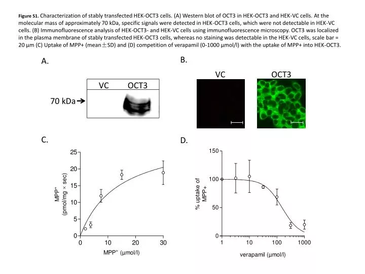
Figure S1. Characterization of stably transfected HEK-OCT3 cells. (A) Western blot of OCT3 in HEK-OCT3 and HEK-VC cells. At the molecular mass of approximately 70 kDa, specific signals were detected in HEK-OCT3 cells, which were not detectable in HEK-VC cells. (B) Immunofluorescence analysis of HEK-OCT3- and HEK-VC cells using immunofluorescence microscopy. OCT3 was localized in the plasma membrane of stably transfected HEK-OCT3 cells, whereas no staining was detectable in the HEK-VC cells, scale bar = 20 µm (C) Uptake of MPP+ (mean±SD) and (D) competition of verapamil (0-1000 µmol/l) with the uptake of MPP+ into HEK-OCT3.
Characterization of stably transfected HEK-OCT3 cells. (A) Western blot of OCT3 in HEK-OCT3 and HEK-VC cells. At the molecular mass of approximately 70 kDa, specific signals were detected in HEK-OCT3 cells, which were not detectable in HEK-VC cells. (B) Immunofluorescence analysis of HEK-OCT3- and HEK-VC cells using immunofluorescence microscopy. OCT3 was localized in the plasma membrane of stably transfected HEK-OCT3 cells, whereas no staining was detectable in the HEK-VC cells, scale bar = 20 µm (C) Uptake of MPP+ (mean±SD) and (D) competition of verapamil (0-1000 µmol/l) with the uptake of MPP+ into HEK-OCT3.