Download

1 / 62
680 likes | 1.15k Views
Neurosurgery. Outline. A & P Pathology Diagnostics/Pre-operative Testing Medications/Anesthesia Positioning/Prepping/Draping Supplies/Instrumentation/Equipment Dressings/Drains/Post-op Care

E N D
Outline • A & P • Pathology • Diagnostics/Pre-operative Testing • Medications/Anesthesia • Positioning/Prepping/Draping • Supplies/Instrumentation/Equipment • Dressings/Drains/Post-op Care • Procedures: Carpal Tunnel Release, Craniotomy, Cervical Discectomy, Lumbar Discectomy, Ventroperitoneal Shunt
Nervous System • Functions: • Senses changes in environment • Interprets changes • Stimulates movement to respond to these changes
Organization of the Nervous System • Two systems: 1. CNS Central Nervous System • Two major parts: Brain and Spinal Cord 2. PNS Peripheral Nervous System • Everything else
Peripheral Nervous System • Two major parts: • Afferent Nervous System • Sensory neurons take info from PNS to CNS • Efferent Nervous System • Motor neurons take info from CNS to PNS
Efferent Nervous System • Motor nervous system • 2 parts: • Somatic Nervous System • Skeletal muscle control • Conscious control • Autonomic Nervous System • Cardiac muscle, smooth muscle, and glands • Unconscious control • Has 2 divisions: • Sympathetic Division • Parasympathetic Division
Autonomic Nervous System • Sympathetic vs. Parasympathetic • Controlled by hypothalamus and medulla oblongata • Both are different nerves going to the same effector or target • Are antagonistic • Parasympathetic = decreased skeletal blood flow, increased organ blood flow (quietly eating) • Sympathetic = increased skeletal blood flow, decreased organ blood flow (eatus interruptus by a bear!) Also called fight or flight; prepares body for emergencies
Spinal Cord • Functions: • Info to and from the brain • Integration of reflexes • Location: • Begins at foramen magnum and extends to 2nd lumbar • About 16-18” in length
Spinal Cord Support Structures • Vertebra • 33 total • 7 cervical • 12 thoracic • 5 lumbar • Sacrum formed by 5 fused bones • Coccyx formed by 4 fused bones
Intervertebral Disks • Separate vertebrae • Outer layer is tough and called the annulus fibrosis • Inner core is soft and called the nucleus pulposus • Bear stress incurred with body weight and when lifting
Spinal Cord Support Structures • Meninges • Between vertebra & spinal cord • Epidural space between vertebra and dura mater • Dura Mater outermost layer extends to S-2 • Subdural space between dura mater and arachnoid • Arachnoid extends to S-2 • Subarachnoid space contains CSF • Pia Mater adheres directly to spinal cord and extends to L-2 • Meninges also cover brain/continuous layer/difference epidural space not present
Spinal Nerves • 31 pair • Names and numbers depend on where enter and exit • Each has a ventral and dorsal root • Ventral root = motor • Dorsal root = sensory • 8 cervical • 12 thoracic • 5 lumbar • 5 sacral • 1 coccygeal
Brain • Protected by the cranium or skull
Brain • 4 major parts: • Brain stem • Diencephalon • Cerebellum • Cerebrum Weight about 3 lbs.
Support Structures of the Brain 1. Meninges • Continuous layer with spinal cord • NO epidural space
Support Structures of the Brain 2. Cerebrospinal fluid (CSF) • About 800ml produced each day by the choroid plexus, a specialized set of capillaries • Circulates inside subarachnoid space through central canal of spinal cord and the ventricles of the brain • Reabsorbed in arachnoid villus found in the parietal lobe • Functions as a shock absorber and circulates nutrients
Support Structures of the Brain • Blood Brain Barrier • Specialized set of capillaries exclusive to the central nervous system • Less permeable than any other capillaries in the body • Advantage = keeps out unwanted chemicals • Disadvantage = difficult to diffuse materials out, hence difficulty in treating conditions such as encephalitis
Brain Stem • 3 parts: • Medulla oblongata • Pons • Midbrain
Medulla oblongata • Contains: • 5 of 12 cranial nerves • Pyramids: function only with motor neurons/a crossing of the spinal nerve impulses • Reflex centers: hiccupping, sneezing, coughing • Vital reflex centers: • Cardiac center – heart rate • Vasoconstrictor center-BP via blood vessel diameter control • Respiratory center - breathing
Pons • Above medulla • Switching point for motor neurons • Respiratory center
Midbrain or Mesencephalon • Above pons • Involuntary eye and head movement in response to auditory stimuli
Diencephalon • 2 parts: • Thalmus • Hypothalmus
Thalmus • Relay center for sensory information • Interprets stimuli for example pain from changes in temperature (hot stove) • 1st level of reasoning occurs here • Recognizes crude touch NOT localized touch
Hypothalmus • Controls large number of subconscious functions • Controls most of Autonomic nervous system • Where endocrine and nervous systems interface • Homeostasis regulation of the body • Controls: body temp, thirst, hunger, sleep and waking habits, psychosomatic disorders, rage and aggression
Cerebellum • 2nd largest part of the brain • Primarily a motor area • Controls skeletal muscles, subconsciously • Receives sensory input from eyes, muscles, joints, and inner ear • Posture, balance, coordination, equilibrium • Muscle sense tells body where other parts are
Cerebrum • Largest part of brain • Motor/sensory/association area • 4 Lobes: frontal, parietal, occipital, temporal • Each controls a specific function be it motor or sensory • Limbic system: controls emotion/functions in cerebral cortex and diencephalon • See page 970 Figure 24-4 in Price
Frontal Memory, abstract thinking, ethics, judgement, emotion, expressive speech, motor Parietal Sensory, receptive speech, written word Temporal Auditory, olfactory Occipital Visual cortex Visual association Cerebrum Lobes’ Function
Cranial Nerves • All originate in the brain stem EXCEPT the 1st and 2nd • Classified as sensory or mixed (sensory and motor) nerves • Directly off of brain • Do not go through the spine • Identified by Roman numerals and names
Cranial Nerves • Olfactory - sense of smell • Optic – sense of sight/vision • Occulomotor – eyeball, eyelid movement (medial, inferior, superior rectus, inferior oblique), pupil constriction, lens accommodation Muscle sense for eyeball • Trochlear – eyeball movement (superior oblique) Muscle sense for eyeball • Trigeminal – masseter muscle control Sensory part has 3 branches: ophthalmic (forehead to corner of eye), maxillary (corner of eye to upper lip/teeth), and mandibular (lower lip/teeth/tongue) All three convey sense of touch, pain and temp changes • Abducens - same as IV eyeball movement (lateral rectus) and eyeball muscle sense FYI: EOM formula LR6(SO4)3
Cranial Nerves • Facial- facial muscles, lacrimal and salivary glands anterior 2/3 of tongue (taste) • Vestibulocochlear -last of totally sensory nerves; has 2 branches: vestibular conveys balance and cochlear which conveys sense of hearing • Glossopharyngeal -salivary gland secretion and posterior 1/3 of tongue • Vagus – internal organ control motor and sensory; originates in medulla and goes down through neck into chest and abdomen • Accessory – controls head and neck movement, speech, and muscle sense for the head • Hypoglossal – tongue muscles: swallowing, speech, muscle sense for tongue
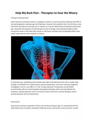
Cervical Spine Pathology • Very serious • Can have severe consequences related to all of the spinal cords’ nerve pathways • Spondylosis is osteophyte or bone spur formation in the spinal canal • Cervical disk extrusion acute or chronic • Treatment errs on the side of caution due to potential extreme consequences of surgical intervention
Thoracic Pathology • Spondylosis • Extrusion of disk
Lumbar Pathology • Spondylosis • Stenosis • Spondylolithesis • Disk extrusion
Neoplasms/Tumors • Two types: • Primary • Originate in nervous tissue or meninges • Secondary • Are metastasized from other parts of the body • May be classified as benign or malignant
Tumors • Benign tumors: • “Craniopharyngiomas, epidermoids, hemangiomas, menigiomas, acoustic neuromas, and pituitary microadenomas” Price, 2004 • Malignant tumors: • “Astrocytes or gliomas” Price, 2004 • Benign usually excisable via craniotomy • Malignant normally cannot be completely removed but efforts are made to remove most
Head Trauma • Includes; • Scalp lacerations, fractures, hematomas (epidural or subdural), and brain injuries
Spinal Cord Trauma • Vertebral Fracture • Vertebral Dislocation • Herniated disk into spinal canal • Laceration from GSW or MVA
Cerebrovascular Disease • #3 cause of death in US • Symptoms reflect ischemia (TIAs) or hemorrhage • Intracranial aneurysm • Arteriovenous malformations • Brain hemorrhage • Stroke or cerebrovascular accident (CVA)
Congenital Pathology • Craniosynotosis “premature closure of the cranial sutures” Price, 2004 • Hydrocephalus result of obstructed CSF flow • Spina bifida
Infection • Abscess • Subdural empyema • Post-op infection
Spinal Cord Tumors • Intramedullary in the spinal cord • Intradural in dura, outside spinal cord • Extradural outside spinal cord Price, 2004
Peripheral Nerve Pathology • Carpal tunnel syndrome - compression of the median nerve • Ulnar nerve compression – compression of ulnar nerve by the ligament of Osborne Price, 2004