Download
1 / 13
130 likes | 271 Views
Evaluation-The Foot and Toes. Ms. Bowman. Anatomy Review-Bones. 26 bones Phalanges-toes; proximal, middle, and distal Metatarsals-5; between phalanges and tarsals Tarsals-calcaneus, talus, navicular , cuboid, 3 cuneiforms Divided into 3 sections

E N D
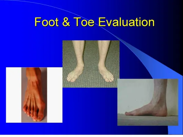
Evaluation-The Foot and Toes Ms. Bowman
Anatomy Review-Bones • 26 bones • Phalanges-toes; proximal, middle, and distal • Metatarsals-5; between phalanges and tarsals • Tarsals-calcaneus, talus, navicular, cuboid, 3 cuneiforms • Divided into 3 sections • Rearfoot-formed by the calcaneus and talus; provides stability and shock absorption during the initial stance phase of gait and serves as a lever arm for the Achilles tendon during plantarflexion • Midfoot-composed of the navicular, three cuneiforms, and cuboid; shock absorbing section of the foot • Forefoot and toes-formed by the 5 metatarsals and 14 phalanges; act as a lever during the preswing phase of gait
Anatomy Review-Articulations and Ligaments • Subtalar Joint-articulation of the calcaneus and talus • Midfoot- • midtarsal joint formed by the articulations of the tarsal bones • Plantar calcaneonavicular ligament (spring liegament) • Forefoot- • Tarsometatarsal joints-junction between the midfoot and forefoot • Intermetatarsal joints-proximal and distal joints • Metatarsophalangeal joints • Interphalangeal joints • Medial Ligaments • Deltoid ligament-composed of 4 ligaments • Posterior tibiotalar ligament • Tibiocalcaneal ligament • Anterior tibiotalar ligament • Tibionavicular ligament • Lateral Ligaments • Anterior talofibular ligament • Calcaneofibularligament • Posterior talofibular ligament
Anatomy Review-Muscles • There are many muscles that act on the foot. • Those that originate and insert in the foot are called intrinsic foot muscles. These directly influence the foot and toes. • Those that originate in the lower leg are called extrinsic foot muscles. These influence motion at the ankle and knee as well as the foot and toes. • If the muscle name begins with extensor, then the muscle’s primary function is extension. • If the muscle name begins with flexor, then the muscle’s primary function is flexion.
History • Location of p!-trauma to intrinsic structures or secondary to compensation for improper lower leg biomechanics • Metatarsal p!-p! that worsens over time can indicate stress fx; p! between the MTs possibly a result of nerve impingement • Great toe p!-localized p! to plantar surface can be indicative of sesamoidfx; p! with flexion or extension can be an indicator for turf toe • Onset and Mechanism of injury • Acute onset- can occur from trauma (fx, ligament sprain, muscle strain) • Insidious onset-result of overuse injuries; may be the result of playing surface, distance and duration of exercise, shoes
Inspection • Analyze gait • Look for • Gross deformity • Edema • Redness • Calluses and blisters (indicates improperly fitting shoes, poor mechanics, or underlying bony or soft tissue dysfunction) • Observe • Alignment of the toes • Toenail integrity (ingrown toenails, subungual hematoma) • Arches • Achilles • Calcaneus • Plantar surface of the foot
Palpation • When palpating, check for deformity, alignment, edema, crepitus, and pain • Medial Structures • 1st Phalange, 1st MT, 1st Cuneiform, Navicular, Talar head Spring Ligament, Calcaneus • Lateral Structures • 5th phalange, 5th MT, Cuboid, Peroneal tubercle, Calcaneus • Dorsal Structures • Rays, Cuneiforms, Navicular, Dome of Talus, Musculature • Plantar Structures • Plantar fascia, MT heads, sesamoid bones of great toe
ROM Testing • ROM should be measured with a goniometer • AROM, PROM, and RROM should be assessed as necessary • Goniometry measurements • Fulcrum-placed over joint • Stationary arm-placed over the proximal (non-moving part of body) • Movement arm-placed over the distal (moving part of body)
Ligamentous Testing • Valgus and Varus stress tests should be used to test the integrity of the MTP and IP joints • Metatarsal gilde test can be used to tests for the integrity of the ligaments connecting the MT • Midtarsal joint glide test can be used to test the integrity of the intertarsal ligaments
Neurological Assessment • Foot and toes are supplied by L4 and S2 nerve roots • Can be assessed by doing a lower quarter screen
Special Tests • Navicular drop test • Tinel’s sign • Long bone compression test • Pencil test