Download
1 / 37
510 likes | 1.14k Views
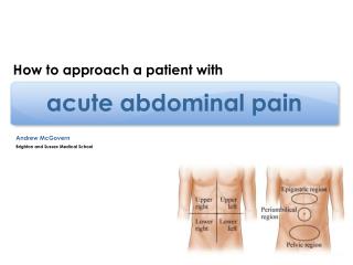
Approach to the patient with acute abdominal pain. Asisst. Prof. Dr.Özlem Tanrıöver Yeditepe University Medical Faculty Department of Family Medicine. Abdominal Anatomy. Four quadrants: Right Upper Quadrant Right Lower Quadrant Left Upper Quadrant

E N D
Approach to the patient with acute abdominal pain Asisst. Prof. Dr.Özlem Tanrıöver Yeditepe University Medical Faculty Department of Family Medicine
Abdominal Anatomy • Four quadrants: • Right Upper Quadrant • Right Lower Quadrant • Left Upper Quadrant • Left Lower Quadrant • Three central areas: • Epigastric • Periumbilical • Suprapubic
Abdominal Anatomy • Right upper quadrant Liver, Head of Pancreas, Kidney and Lung Right lower quadrant Appendix, Ureter, Bladder, Colon, Gonads Left upper quadrant Heart, Spleen, Body of pancreas, Kidney, Stomach, Lung Left lower quadrant Ureter, bladder, colon, gonads
The History and Physical in Perspective • 70% of diagnoses can be made based on history alone. • 90% of diagnoses can be made based on history and physical exam. • Expensive tests often confirm what is found during the history and physical.
Types of Abdominal Pain • Pain from Hollow Viscera • crampy/paroxismal • often poorly localized • related to peristalsis • patient writhing on exam table • Pain from Peritoneal Irritation • steady/constant • often localized • patient lies still with knees up
Key Historical Points - Bowel and Bladder • Nausea, Vomitting, Diarrhea, Constipation • Frank Blood, "Coffee Grounds" Emesis, Black Stools • Urinary Frequency, Urgency, Discomfort
Key Historical Points - Reproductive • Sexual Activity, Contraception, Last Menstrual Period • Always Consider Pregnancy in Reproductive Age Women • Have a Low Threshold for Pregnancy Testing
Gastrointestinal Review of Systems • Trouble swallowing • Heartburn • Loss of appetite • Nausea • Change in bowel habits • Blood in stool • Dark tarry stools • Constipation • Diarrhea • Abdominal pain • Jaundice • Fever or chills
Ask the following questions: • Where is the pain? • Has the pain changed its location since it started? • Do you feel the pain in any part of your body? • How long have you had the pain? • Have you had recurrent episodes of abdominal pain?
Ask the following questions: • Did the pain start suddenly? • Can you describe the pain? Is it sharp, burning, cramping? • Is the pain continuous? • What makes it worse, or better? • Is the pain associated with nausea, vomiting, sweating, constipation, diarrhea, bloody stools, abdominal distention, fever, chills, eating? • If the patient is a woman; • When was your last period?
Abdominal Pain • Location • Other symptoms • Character • Factors that • aggravate or • alleviate • Timing • Environment • Severity
Common causes of acute abdominal pain • Peptic Ulcer Disease • Cancer of Stomach • Pancreas, Colon • Biliary Colic • Acute Cholecystitis • Acute Appendicitis • Acute Diverticulitis • Intestinal Obstruction • Mesenteric Ischemia • Irritable Bowel Syndrome • Inflammatory Bowel Dis. • Hepatitis • Gastroenteritis
Peptic Ulcer Disease or Dispepsia • Ulcer begins in lining of stomach or duodenum. • Helicobacter pylori infection is often present. • Dyspepsia more common ages 20- 29, gastric ulcer in those over 50 and duodenal ulcer ages 30 – 60.
Peptic Ulcer Disease or Dispepsia • Pain is epigastric and may radiate to the back. • Variable, gnawing, burning, boring, aching or hungerlike. • Timing is intermittent. Duodenal ulcer more likely nocturnal. • Food and antacids may relieve duodenal ulcer pain. • Accompaning symptoms include nausea, vomiting, belching, bloating, heartburn and weight loss.
Pancreatitis • Inflammation of the pancreatic tissue often due to gallstones or alcohol abuse. • Pain is epigastric and may radiate to the back. • Pain onset is acute and pain is steady. • Pain may be worse when supine and relieved with leaning forward. • Associated with nausea, vomiting, abdominal distention and fever.
Biliary Colic and Acute Cholecystitis • Due to obstruction of the cystic duct or common bile duct by a gallstone. • Pain is epigastric or right upper quadrant and may radiate to the right scapula or shoulder. • The pain is steady and aching. • Biliary colic may start suddenly and subside then recur whereas cholecystitis is more steady. • Associated with anorexia, nausea, vomiting and fever.
Classic Presentations – Acute Cholecystitis Localized or diffuse RUQ pain Radiation to right scapula Vomitting and constipation Low grade fever
Acute Diverticulitis • Inflammation of a colonic saclike mucosal Outpouching through the colonic muscle. • Pain is in the left lower quadrant. • The pain may begin as cramps then become steady. • It is associate with fever, constipation and sometimes, brief diarrhea.
Acute Appendicitis • Acute distention or obstruction of the appendix. • Pain often begins as poorly localized periumbilical pain followed by right lower quadrant pain. • Becomes more steady and severe with time. • Pain is worse with movement or cough. • Pain is associated with anorexia, nausea, and possibly vomiting which typically follow the onset of pain.
Classic Presentations - Acute Appendicitis • Diffuse periumbilical pain and anorexia early • Pain localizes to RLQ as peritonitis develops • Low grade fever, nausea and vomitting may not be present • Xrays and other tests are often negative
Psoas Sign • This is a test for appendicitis. • Place your hand above the patient's right knee. • Ask the patient to flex the right hip against resistance. • Increased abdominal pain indicates a positive psoas sign.
Obturator Sign • This is a test for appendicitis. • Raise the patient's right leg with the knee flexed. • Rotate the leg internally at the hip. • Increased abdominal pain indicates a positive obturator sign
Rovsing's Sign Tenderness felt in the RLQ when palpation is performed on the left is called Rovsing’s sign and suggests appendicitis.
Classic Presentations - Acute Renal Colic • Severe flank pain • Radiation to groin • Vomitting and urinary symptoms • Blood in the urine
Inflammatory Bowel DiseaseUlcerative Colitis • Inflammation of colon • Soft bloody stools • Insidious onset • Associated with crampy lower or generalized abdominal pain, anorexia, weakness and fever • Often begins in young people
Inflammatory Bowel DiseaseCrohn’s Disease • Chronic inflammation of the bowel wall, typically involving the terminal ileum and/or proximal colon • Stools loose but not as bloody • Insidious onset • Associated with crampy periumbilical or right lower quadrant pain with anorexia, low fever and/or weight loss. • Perianal or perirectal abcesses and fistulas common • May begin in youth or later.
Physical Examination • Pelvic • Genital • Rectal exam on every patient • with severe abdominal pain
Laboratory Evaluation • CBC, Urine Analysis, Electrolytes • Urine and serum pregnancy test in all women of reproductive age with lower abdominal pain • Liver Function Tests , amylase/lipase on all with upper abdominal pain
Radiographic Evaluation • Plain radiograph • upright and supine abdomen and chest x-ray • Ultrasound on patients with biliary and pelvic symptoms • CT Abdomen and Pelvis • evaluates vasculature, inflammation and solid organs
The differentialdiagnosis • Acute Cholecystitis • cystic duct obstructed, RUQ pain ? R scapula • LFTS, amylase • Acute Appendicitis • anorexia, N/V and vague periumbilical pain • 6-8 hrs pain migrates to RLQ, fever • Progresses to localized peritoneal irritation • CT useful in diagnosis
The differential • Pancreatits • Acute Diverticulitis • most commonly in sigmoid colon • symptoms related to inflammation or obstruction • CT useful early to r/o absess, Endoscopy contraindicated ? wait 4-6 wks • Rx bowel rest, IV abx, surgery for failures
Pregnancy appendicitis, cholecystitis, pyelonephritis, adnexal problems (ovarian torsion, ovarian cyst rupture) appendicitis 7/1000 pregnancies 3% fetal loss with surgery, but 20% with perforated appendix
Summary • Obtain detailed history • Careful exam • Consider patient circumstances (diabetes, age, previous ab surgery) • Early thorough work-up (labs/x-rays) • Frequent evaluation of progression
Rebound Tenderness • This is a test for peritoneal irritation. • Warn the patient what you are about to do. • Press deeply on the abdomen with your hand. • After a moment, quickly release pressure. • If it hurts more when you release, the patient has rebound tenderness
Psoas Sign • This is a test for appendicitis. • Place your hand above the patient's right knee. • Ask the patient to flex the right hip against resistance. • Increased abdominal pain indicates a positive psoas sign.
Obturator Sign • This is a test for appendicitis. • Raise the patient's right leg with the knee flexed. • Rotate the leg internally at the hip. • Increased abdominal pain indicates a positive obturator sign
Rovsing's Sign Tenderness felt in the RLQ when palpation is performed on the left is called Rovsing’s sign and suggests appendicitis.