Download
1 / 20
210 likes | 390 Views
Medipix Collaboration. Large scale x-ray images taken with the Medipix1 chip. Karl-Friedrich Pfeiffer Physikalisches Institut, Abt. 4 Universität Erlangen-Nürnberg Workgroup SPOC *. * S ingle P h O ton C ounting. Overview. The Medipix1 chip Experimental setup

E N D
Medipix Collaboration Large scale x-ray images taken with the Medipix1 chip • Karl-Friedrich Pfeiffer • Physikalisches Institut, Abt. 4 • Universität Erlangen-Nürnberg • Workgroup SPOC* *Single PhOton Counting
Overview • The Medipix1 chip • Experimental setup • Large Images 1: 'Move and Tile'Measurement and Simulation • Large Images 2: 'Tile and Move'Measurement and Simulation • DQE Measurements • Summary
The Medipix1 chip • Pixelated solid state hybrid detector for single photon counting • 64 x 64 Pixel, size 170mm Þ active area 10.88 x 10.88 mm2 • Semiconductor sensor layer bump-bonded to the pixelated read-out electronics chip • Sensor layer: e.g. 300mm Si, but other materials (GaAs, Cd(Z)Te...) can be used as well • Developed within the framework of the Medipix1 collaboration
Computer 1: main control Computer 2: data acquisition X-ray generator MUROS1 board & Medipix1 chip Translation stages Experimental setup • Complete imaging process controlled by computer 1 • Data acquisition done with Medisoft software (developed in the Medipix collaboration) • (Semi-)automated processing of acquired data
Large Images 1: 'Move and Tile' • Make single exposure with single detector • Move detector to next position • Make another exposure • Repeat until the whole area is covered • Merge all single exposures to get one large tiled image
Large Images 1: Measurement 5x5 single exposures, 10 pixels overlap
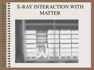
Contrast ratio measurement -simulation: 0.996 0.978 Comparison Measurement - Simulation Object: „Siemensstern“ 0.05mm Pb in 2mm PMMA Ø 4.5cm • Contrast Pb -Air:0.965 • Contrast Pb-PMMA:0.941 • Contrast Pb -Air:0.969 • Contrast Pb-PMMA:0.962
Large Images 2: 'Tile and Move' • Tile several detectors to make an array • Take large picture (with gaps) • Move array to next position • Take another picture • Repeat to cover all gaps • Merge pictures to get one large image without gaps and with high photon statistics
3 images, 4 x 4 tiles each Large Images 2: Measurement
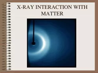
Contrast ratio measurement -simulation: 0.988 0.964 Comparison Measurement - Simulation Object: „Siemensstern“ 0.05mm Pb in 2mm PMMA Ø 4.5cm • Contrast Pb -Air:0.954 • Contrast Pb-PMMA:0.928 • Contrast Pb -Air:0.966 • Contrast Pb-PMMA:0.963
Improved 'Tile and Move' If it is possible to tile several detecors without gaps (e.g. with the Medipix2 chip): Þ Tile detectors to get ’detector bars': two pictures are sufficient to cover all gaps Þ even faster & less dose!
DQE Definition • DQE: Detective Quantum Efficiency • SNR: Signal-to-Noise-Ratio • in: SNR of incoming radiation field (~ N ) • out: SNR of the image • f = f(x,y): Spatial frequency
Calculation of the DQE : System gain factor MTF : Modulation Transfer Function NPS : Noise Power Spectrum <E> : Mean photon energy 0 : Incoming energy fluence
15.5cm DQE Measurement • MTF(f) measurement:line pair phantom • NPSout measurement:flat field images • ... some lenghty calculations ... Þ DQE(f)
Summary • There are basically two ways to get large field images: 1) 'Move and Tile': single detector, flexible, but takes quite a long time and high dose Þ 'Lab Method' 2) 'Tile and Move': quite fast, but requires several detectors Þ 'Standard Imaging Method' • The MTF and DQE of a Medipix1 chip with a 300mm Si sensor layer was determined • We are looking forward to the Medipix2 chip
Thanks to: • Prof. G. Anton1 • Ch. Bert1 & D. Niederlöhner1 • J. Giersch1 • M. Hoheisel2 & L. Bätz2 (for the DQE calculations) • Medipix Collaboration • B. Mikulec3 1Universität Erlangen 2Siemens 3CERN
Phantoms used: Siemensstern Line pair phantom