Download

1 / 12
120 likes | 290 Views
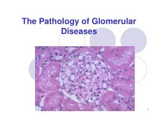
The Physiology of the Glomerular Tuft. Structure. The glomerulus consists of a capillary tuft that is surrounded by Bowman's capsule, which passes the filtered primary urine to the tubular system.

E N D
Structure • The glomerulus consists of a capillary tuft that is surrounded by Bowman's capsule, which passes the filtered primary urine to the tubular system. • It is a network held together by connective tissue and composed of three distinct cell types: endothelial cells, mesangial cells, and glomerular visceral epithelial cells (podocytes). • Glomerular filtration barrier is a trilaminar structure composed of a fenestrated endothelium, the hydrated meshwork of the glomerular basement membrane (GBM), and the filtration slit formed by pedicel interdigitation.
Mesangial Cells • There are two distinct populations of mesangial cells. • The first population makes up about 85% to 95% of resident mesangial cells and has a network of contractile elements, such as actin, myosin, and tropomyosin. • The second population exhibit features of monocytes/macrophages and are derived from bone marrow.
The covering of the capillaries by the GBM is partial. • Mesangial cells account for the completion of the capillary covering by forming loops that completely encircle the capillaries. • Those contacts establish a biomechanical unit, with the basement membrane serving as the effector site and mesangial cells as the contractile motor. • These contacts permit the mesangial cells to support the mesangium and regulate the capillary surface area and glomerular volume, influencing glomerular hemodynamics. • Therefore, mesangial cells can modulate GFR by changing the capillary surface area and redistributing blood volume in glomerular capillaries through the actions of mesangial loops.
Contraction of a mesangial cell occurs in response to hormones, vasoactive compounds, and growth factors through G protein–associated receptors that activate phospholipase C. • Hormones using the cyclic AMP and cyclic GMP cascades relax mesangial cells through direct effects or through antagonism of contracting substances.
Glomerular Filter • Endothelial Cell • The glomerular endothelial fenestrae may be covered by a diaphragm formed by a thick cell coat (glycocalyx). • This layer is a matrix-like gel composed of proteoglycans, with negatively charged as well as neutral glycosaminoglycans, glycoproteins, and plasma proteins carrying a negative charge. • Thus, the glycocalyx is an initial charge barrier in the glomerular filter and function as a size-selective barrier.
Glomerular Basement Membrane • The GBM is an especially thick membrane that lies between the endothelial cells and the visceral epithelium (podocytes). • The main components of the GBM are triple-helical type IV collagen, proteoglycans, laminin, and entactin.
Glomerular Visceral Epithelial Cells (Podocytes) • Podocytes can be divided into three distinct functional segments: cell body, primary processes, and secondary processes, also known as foot processes or pedicels. • These cells are located at the external surfaces of glomerular capillaries, which they cover with pedicels.
The covering of capillaries is achieved by interdigitation of pedicels derived from adjacent podocytes, forming between them filtration slits that are bridged by a specialized cell junction, called slit diaphragms, which represent the last filtration barrier to proteins. • The cell body and primary processes are mainly constituted by microtubules and intermediate filaments, whereas pedicels contain a dense network of actin microfilament bundles associated with myosin II, α-actin, talin, and vinculin, forming a complex contractile apparatus.
Podocytes have all the necessary elements to generate tensile strength and stabilize glomerular architecture by counteracting the hydrostatic forces causing distentions of glomerular capillaries. • Podocytes also have receptors for several contracting and relaxing factors.
The slit diaphragm not only provides a structural element, serving as a static molecular sieve, but also is a dynamic multifunctional protein complex that functions as a signalling platform for the podocyte. • Podocytes seem to play a crucial role in the development of the entire glomerular tuft, mainly through production of angiogenic factors. • During glomerular development, they express vascular endothelial growth factor-A (VEGF-A), whereas the endothelial and mesangial cells express the VEGF receptors.