Download

1 / 31
320 likes | 737 Views
Liver. Objectives Location of the liver Indentify the surfaces of the liver and their anatomical relations Porta Hepatis Anatomical Divisions Of the Liver Peritoneal attachments of the Liver Blood Supply of the Liver Gall bladder its relations and blood supply. Liver

E N D
Objectives • Location of the liver • Indentify the surfaces of the liver and their anatomical relations • PortaHepatis • Anatomical Divisions Of the Liver • Peritoneal attachments of the Liver • Blood Supply of the Liver • Gall bladder its relations and blood supply
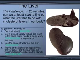
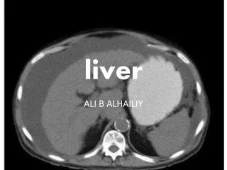
Liver • The liver is the largest visceral organ in the body and is primarily in the right hypochondrium and epigastric region, extending into the left hypochondrium . • Surfaces of the liver include: • a diaphragmatic surface • a visceral surface in the inferior direction .
The diaphragmatic surface of the liver, lies against the inferior surface of the diaphragm . • Associated with it are : • the subphrenic recess • the hepatorenal recess . • The falciform ligament ascends to the liver from the umbilicus and bears the ligamentumteres in its free border.
Liver Falciform Ligament Stomach Greater Omentum Gallbladder Small Intestine
The visceral surface of the liver is covered with visceral peritoneum except in the fossa for the gallbladder and at the portahepatis (gateway to the liver)and structures related to it include: • the right anterior part of the stomach; • the superior part of the duodenum; • the lesser omentum; • the gallbladder; • the right colic flexure; • the right transverse colon; • the right kidney; • the right suprarenal gland.
The PortaHepatis • The portahepatis serves as the point of entry into the liver for the hepatic arteries and the portal vein, and the exit point for the hepatic ducts .
The liver is divided into right and left lobes by fossae for the gallbladder and the inferior vena cava . • The right lobe of liver is the largest lobe, whereas the left lobe of liver is smaller. • The quadrate and caudate lobes are described as arising from the right lobe of liver.
The quadrate lobe is bounded: • on the left by the fissure for ligamentumteres • on the right by the fossa for the gallbladder. • The caudate lobe is bounded • on the left by the fissure for the ligamentumvenosum on the right by the groove for the inferior vena cava. Functionally, it is separate from the right and the left lobes of the liver.
Peritoneal attachments • The falciform ligament • It attaches to the anterior abdominal wall . • The round ligament (ligamentumtereshepatis) is a fibrous cord resulting from the obliteration of the umbilical vein. It is found at the lower free border of falciform ligament. Except for a small area of the liver against the diaphragm (the bare area), the liver is almost completely surrounded by visceral peritoneum . The bare area of the liver is a part of the liver on the diaphragmatic surface where there is no intervening peritoneum between the liver and the diaphragm • The coronary ligament: • anterior coronary ligament; • posterior coronary ligament; chc • Where the coronary ligaments come together laterally, they form the right and left triangular ligaments. • Additional folds of peritoneum connect the liver to the stomach and duodenum respectively. • hepatogastric ligament • hepatoduodenal ligament
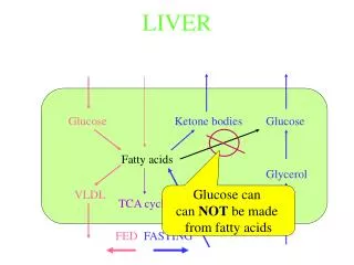
Blood Vessels of the Liver • The liver has a dual blood supply (afferent vessels): • a dominant venous source and a lesser arterial one . The portal vein brings 75 to 80% of the blood to the liver. Portal blood, which contains more oxygen than blood returning to the heart from the systemic circuit, sustains the liver parenchyma (liver cells or hepatocytes)
The portal vein is the final common pathway for the transport of venous blood from the spleen, pancreas, gallbladder, and the abdominal part of the gastrointestinal tract. • It is formed by the union of : • splenic vein • superior mesenteric vein posterior to the neck of the pancreas at the level of vertebra LII .
The hepatic artery, • Accounts for only 20 to 25% of blood received by the liver, is distributed initially to non-parenchymal structures, particularly the intrahepatic bile ducts. • The hepatic artery, a branch of the celiac trunk, may be divided into: • the common hepatic artery, • the hepatic artery proper, from the origin of the gastroduodenal artery . • The hepatic veins drain into the inferior vena cava
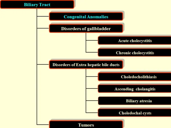
The gallbladder is a pear-shaped sac lying on the visceral surface of the right lobe of the liver in a fossa between the right and quadrate lobes . • It has: • fundus of gallbladder • body of gallbladder • neck of gallbladder. • The gallbladder receives, concentrates, and stores bile from the liver. • The gall-bladder is supplied by the cystic artery (a branch usually of the right hepatic artery