Download

1 / 96
970 likes | 1.16k Views
Explore Raynaud's phenomenon, Raynaud's disease, Erythromelalgia, Livedo Reticularis, Vasculitis, Purpura, and Thrombocytopenic Purpura. Learn about causes, symptoms, treatments & more.

E N D
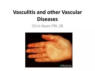
Raynaud’s Phenomenon • Intermittent constriction of the small digital arteries and arterioles • Persistently cyanotic and painful • Aggravated by cold weather • Young middle aged women • Assoc c scleroderma, dermatomyositis, LE, Mixed connective tissue diseases, Sjögren's RA, and paroxysmal hemoglobinuria.
Raynaud’s Phenomenon • Scleroderma is the underlying condition for more than half of the patients • Maybe caused by medications, ie bleomycin • Simple noninvasive tests along with other clinical findings are helpful
Raynaud’s Disease • Primary disorder of cold sensitivity • Pallor, cyanosis, hyperemia, and numbness of the fingers • Precipitated by cold. • Present for 2 years with out associated disease finding • Good prognosis
Etiology ofRaynaud’s Disease/phenomenon • Multifactorial. • Increase alpha-2 sympathetic receptor activity on vessels. • Endothelia dysfunction • Deficiency in calcitonin gene related protein • Central thermoregulatory defects
TX of Raynaud’s Disease/phenomenon • Treatment includes avoidance of the aggravating factor, ie cold. (not just hands) • Vasodilating drugs, nifedipine, 10-20 mg tid; prazosin 1-3 mg tid • Nitroglycerin 2% local application • Sympathectomy in severe disease
Erythromelalgia • Aka erythermalgia and acromelalgia • Rare form of paroxysmal vasodilation affects the feet with burning. Infrequently upper ext. • Burning may last from minutes to days • Triggered by increase in environmental temperature • May be secondary to myeloproliferative disease such as polycythemia vera, TTP
Pathophysiology is poorly understood • Responds to treatment of primary disorders • Cold water immersion • Serotonin antagonists
Mottled or reticulated pink/reddish/blue discoloration Assoc c LE, DM, scleroderma, RA Side effect of amantadine Livedo Reticularis
Necrotizing livedo reticularis • Assoc. with cutaneous nodules and ulcerations • Results from cholesterol emboli in severe atherosclerotic disease • Sneddon’s syndrome, association of LR with the development of a cerebrovascular lesion
Livedoid Vasculitis • Aka - atrophie blanche • Aka - PURPLE (Painful purpuric ulcer with reticular pattern of the lower extremity) • Characterized clinically by early, focal, painful purpuric lesions of the lower extremities that frequently ulcerate and slowly heal • Mostly represents an idiopathic disorder but may be assoc. with systemic disease
Treatment • Low Dosage of Aspirin 325mg qd • Nifedipine 10mg TID • Pentoxifylline 400mg BID-TID
Marshall-White Syndrome “Bier’s Spots” • Marbled mottling produced in the forearm and hand with use of a tight sphgmomanometer • Consist of Bier’s spot and is associated with insomnia and tachycardia • White middle age men
Purpura • Multifocal extravasation of blood into the skin or mucous membrane • Petechiae <3mm • Ecchymosis – deeper and more extensive interstitial hemorrhage • Vibices (vibex) – Linear • Hematoma – pool-like collection, usually walled off by facial planes
Purpura • May result from hyper or hypocoagulable states, vascular dysfunction, and extravascular causes • Complete blood count • PT and PTT • Family history • medications
Thrombocytopenic Purpura • Three Large Categories: • Accelerated platelet destruction, immunologic and nonimmunologic • Deficient platelet production • Unknown pathogenesis
Idiopathic Thrombocytopenic Purpura • Aka autoimmune thrombocytopenic purpura • Aka Werlhof’s disease • Characterized by acute or gradual onset of petechiae or ecchymosis in the skin and mm • Bleeding occurs when platelet count drops below 50,000 • Risk greatly increased for serious hemorrhage when count goes below 10,000
Idiopathic Thrombocytopenic Purpura • Acute variety occurs in children following season viral illness in 50% of the patient. • Lag between illness and onset of purpura is 2 weeks • Most cases resolve spontaneously with minimal therapy • Chronic case may result in death.
Idiopathic Thrombocytopenic Purpura • Chronic form most often occurs in adults • Evaluate patient with Tc99M radionuclide scan to look for accessory spleen • Idiopathic Thrombocytopenic Purpura is the result of platelet injury by antibodies of the IgG class • Treatment include Splenectomy, systemic corticosteroid, IVIg
These are the kidneys from a case of idiopathic thrombocytopenic purpura. Petechiae are found throughout the renal parenchyma.
Drug-Induced Thrombocytopenia • Drug induced antiplatelet antibodies • May be caused by sulfonamides, digoxin, quinine, quinidine, PCN, furosemide, Lidocaine, methyldopa • Remove the offending agent • corticosteroids
Thrombotic Thrombocytopenic Purpura • Aka Moschcowitz’s syndrome • Pentad of thrombocytopenia, hemolytic anemia, renal abnormalities, fever, CNS disturbance. • Delay in diagnosis may lead to a mortality rate as high as 90% • Exchange plasmapheresis, 3 to 5 liters of plasma for 4 to 10 days • Splenectomy may be required
Thrombotic Thrombocytopenic Purpura • Positive histologic diagnosis requires gingival biopsies looking for subendothelial hyaline deposits • Exchange plasmapheresis is required for treatment. 80% patient survive if treatment is instituted.
Dysproteinemic Purpura • Aka Nonthrombocytopenic purpura, purpura cryoglobulinemica, cryofibrinogenemia • Abnormal serum proteins behaving as cryoglobulins and cryofibrinogens • Purpura most apt to occur on exposed surfaces after cold exposure
Occurs most frequently in multiple myeloma and macroglobulinemia of monoclonal IgM, IgG, or Bence Jones cryoglobulin. • Purpura tends to be chronic • Tx with plasmapheresis, systemic steroids, and immunosuppressors.
Purpura Hyperglobulinemica • Aka Waldenström's hyperglobulinemic purpura • Consist of episodic showers of petechiae occurring on all parts of body, most profusely on the lower extremities • Diffuse peppery distribution, resembling Schamberg’s • Induced or aggravated by prolonged walking • Most useful lab test is protein electrophoresis
Purpura Hyperglobulinemica • Hyperglobulinemic purpura occurs most commonly in women. • Frequently seen with Hepatitis C and Sjögren's syndrome, keratoconjunctivitis sicca, RA • Histologically: dermal vessels with perivascular infiltrate of mononuclear cells. • Benign and chronic course. May be assoc. with connective tissue diseases. • Steroids are usually not of benefit
Waldenström's Macroglobulinemia • Bleeding from mucous membrane of the mouth and nose, lymphadenopathy, hepatomegaly, retina hemorrhage, and RARELY purpura • Perivascular infiltrate containing lymphocytes and neutrophils and eosinophils
Waldenström's Macroglobulinemia • Plasmapheresis until adequate dose of chlorambucil is administered. • Cyclophosphamide and corticosteroids are treatment options as well
Drug- and Food Induced Purpura • Drug induced purpura may occur without platelet destruction. • Cocaine induced thrombosis with infarctive skin lesions is assoc. with skin popping. • Rumpel-Leede sign: distal shower of petechiae that occurs immediately after the release of pressure from a tourniquet release. Associated with capillary fragility. • Topical EMLA can induce purpura in 30m.
Solar Purpura • Large, sharply outlined 1-5 cm dark purplish red ecchymoses on dorsum of the forearm • Less frequently, back of the hand
Purpura Fulminans • Aka purpura gangrenosa • Severe, rapidly fatal reaction occurring most commonly in children after infectious illness • Sudden appearance of large ecchymotic areas, esp. prominent over the extremities, progressing to acral hemorrhagic skin necrosis is characteristic • Usually follows some acute infectious disease such as scarlet fever, strep pharyngitis, and meningococcal meningitis, varicella.
Purpura Fulminans • Assoc. with Protein C or S deficiency in Neonates • Management is supportive • Protein C replacement if protein C deficiency is present • Fresh frozen plasma maybe useful • Amputations and deaths continue in severest forms of the disease
Disseminated Intravascular Coagulation • Up to 2/3 of DIC patients have skin lesions • Minute, widespread petechiae, ecchymoses, ischemic necrosis of the skin and hemorrhage bullae. • Elevated PT and PTT, fibrin degradation products • Decrease platelets, decreased fibrinogen