Download

1 / 29
300 likes | 477 Views
Ulcer Foot By Dr R.N.M. Francis M.S Prof of surgery SBMC. Defenition A breach in continuity of skin or epithelium, due to molecular death of tissue. Parts of an ulcer Margin Edge Floor Discharge Base.

E N D
Ulcer Foot By Dr R.N.M. Francis M.S Prof of surgery SBMC
Defenition A breach in continuity of skin or epithelium, due to molecular death of tissue. Parts of an ulcer Margin Edge Floor Discharge Base
Margin of the ulcer denotes the junction between the normal and the ulcerated area. It gives the shape of the ulcer: Round Oval Irregular
Floor of the ulcer • The exposed part of the ulcer is called the floor • The floor may be covered by: • Red granulation tissue --------- healing ulcer • Unhealthy granulation tissue • Slough ---------- Infected • Wash leather ---------- syphilis • GRANULATION TISSUE • Proliferation of new capillaries and fibroblasts intermingled with RBC and WBC with thin fibrin cover over it .
Edge of an ulcer It is the part of the ulcer between the floor and the margin. It denotes the nature of the ulcer: Sloping --------- healing Everted ---------- malignancy Undermined ------tuberculosis Punched out ------ penetrating
Base of an ulcer Base is the structure on which the ulcer lies. It is a palpatory finding. Marked induration is a feature of malignancy.
Discharge in an ulcer Serous ------- healing Purulant ------ infected Bloody --------- neoplastic Serosanguinous ----- infected Greenish ------- Pseudomonas infected
Examination of an ulcer • General survey: • Build of the patient • Evidence of any systemic disorder • 2) Local examination : • Inspection • Palpation • 3)Regional examination: • a) Examination of lymph nodes • b) Examination for vascular insuffiency ---- peripheral pulses • c) Examination for varicose veins • d) Examination for nerve lesion
Types of ulcers • Ulcers can be grouped depending upon • Nature of progress • Healing ulcer • Spreading or active ulcer • Callous ulcer • Zones in the margin of healing ulcer • Red zone : Healing zone and reflects granulation tissue. • White zone: Denotes area of fibrous tissue reaction on the skin side • Blue zone: Junction between the two.
b) Nature of pathology Nonspecific ulcers Specific ulcers Malignant ulcers
Nonspecific ulcers No specific aetiological cause Traumatic: mechanical, physical, chemical, radiation. Arterial Venous Trophic Tropical ulcer With associated diseases: anaemia, nephritis, diabetes, rhematoid arthritis. Miscellaneous: Bazin’s ulcer Mortorell’s ulcer Meleney’s ulcer
Specific ulcers Caused by specific aetiological factors Produces typical features for that aetiology Types: Tuberculous Syphilis Actinomycosis Meleney’s ulcer Hemolytic strepococcal gangrene Meleney’s ulcer
Malignant ulcers Epithelioma Rodent ulcer Melanoma
Non specific ulcers • Ischaemic ulcers • Due to poor blood supply • Develop over limbs • Over pressure areas • Superficial, later become deep • Painful • Can be multiple
Venous ulcers • Complication of varicose veins and DVT • Due to ambulatory venous hypertension • Seen in the lower third of medial aspect of leg because of the presence of direct perforating veins which transmit the pressure changes directly to the superficial system.
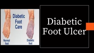
Trophic ulcers (Penetrating ulcers) Seen in: Neurological cases Hansen’s disease Diabetes Common sites: Heel Ball of foot Sacrococcygeal region Features: Deep ulcers Base may be formed by underlying bone Punched out edges Foul smelling slough Surrounding insensitivity
Tropical ulcers • These ulcers are sometimes seen in tropical countries. • They are also called as Delhi boil, Baghdad sore. • They are thought to be due to Vincent’s organism. • It starts as an indurated papule on exposed surface. • Leads to formation of an indolent ulcer. • Leaves back an ugly and pigmented scar.
Tuberculous ulcer • The edge of the ulcer is undermined. • Pale granulation tissue in the floor. • Serous discharge. • It results secondary to caseous lymph nodes.
Marjolins ulcer Squamous cell carcinoma developing in a scar tissue or chronic ulcer. Everted edges Indurated base Bleeding on touch Regional lymph nodes are not involved. Poor response to radiotherapy
Footballer’s ulcer • Also called as traumatic ulcer. • Occurs on shin of tibia. • If not treated can become indolent and adherent to bone. • Usually acquired during game of football.
Investigations 1) Lab investigatios Urine routine Blood urea Blood sugar 2) Discharge for culture and sensitivity. 3) Staining of the discharge for AFB. 4) Wedge biopsy. 5) X-ray
Treatment Conservative Rest to the part Avoid local irritation Improve the nutrition: Protein supplementation Vitamin supplementation Blood transfusion Appropriate antibiotics Treat the cause
Local methods of taking care of the ulcer 1) Separation of slough Hypochlorite solution 0.5% silver nitrate Normal saline soaks 2) Local coverage of the ulcer Amnion Gauze impregnated with antibiotics --- Sofra tulle
3) Excessive granulation Excision Curettage Application of copper sulphate crystals Surgical methods that may be employed Excision of the ulcer and grafting Covering the area with SSG
Requisites for an ideal dressing • Should maintain high humidity between wound and the dressing. • Remove excess exudates. • Permit gaseous exchange. • Impermeable to microorganisms • Allow easy removal • Cost effective
Some of the local wound care modalities Agent Composition Function Commercial names Polymer films Polyurethane Allows water vapour Opsite, tagaderm permeation Hydrocolloids Hydrophilic colloid Impermeable to fluids Intrasite particles & bacteria Alginates Seaweed polymer Absorbs exudates Algisorb non-adherent Medicated Soframycin Topical antibiotic Sofratulle Gauze Bacitracin RhPDGF Acts through Stimulates angiogenesis Plermin RhEGF tyrosinekinase Stimulates Regen- D receptor epithelialisation
Diabetic ulcer Control the sugar Perform culture/ sensitivity Desloughing Antibiotics Local amputation/ disarticulation
Venous ulcers Elevation Compression stockings Treat varicose veins
Trophic ulcers Protection and soft padding. MCR shoes to redistribute the pressure points. Desloughing and debridement are carried out. Amputation or disarticulation of bone involved.