Download
1 / 36
390 likes | 1.05k Views
Radiographic Imaging in Inflammatory Bowel Disease. Ed Barnes, MIV August 28, 2008. Inflammatory Bowel Disease. Crohn’s Disease (CD) Ulcerative Colitis (UC). Brief Review. CD and UC typically manifest in young adolescents and young adults (2 nd to 4 th decade of life)

E N D
Radiographic Imaging in Inflammatory Bowel Disease Ed Barnes, MIV August 28, 2008
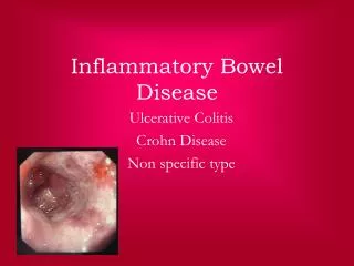
Inflammatory Bowel Disease • Crohn’s Disease (CD) • Ulcerative Colitis (UC)
Brief Review • CD and UC typically manifest in young adolescents and young adults (2nd to 4th decade of life) • No differences in prevalence between males and females
Brief Review • Intestinal inflammation is typically limited to the colon in patients with UC • CD can involve the entire GI tract, from mouth to anus
Clinical Presentation • 85% of patients with CD and 70% of patients with UC report >5 bowel movements per day during acute phases • Acute phases of UC almost always present with bloody diarrhea • Bloody diarrhea in CD indicates extensive rectal and/or sigmoid colon involvement, and is more rare
Extraintestinal Manifestations • Cutaneous lesions • Orofacial lesions (more common in CD) • Opthalmologic manifestations • Arthropathies • Hepatobiliary manifestations • Fatty liver degeneration • Primary sclerosing cholangitis • Cholelithiasis • Granulomatous hepatitis
Diagnosis and Differentiation • Diagnosis of IBD and differentiation between the two subtypes utilizes many modalities • Clinical Impression • Radiologic Findings • Endoscopic Studies • Histologic Criteria
Endoscopy • Endoscopic examination has long been a foundation in the diagnosis of IBD • Allows for direct visualization of the mucosa • Allows for systematic biopsy retrieval
Endoscopy Nikolaus and Schreiber
Plain Abdominal Film • Typically indication is limited to emergency circumstances such as abdominal distension • Fastest diagnostic procedure in patients with acute abdominal pain
Abdominal Plain Film • Provides detection of free intraperitoneal air if perforation has occurred • Air fluid levels can easily be seen • May reveal small bowel obstruction, large bowel dilation, or toxic megacolon
Upper GI Series • Upper GI Series, as well as Small Bowel Follow Through, use barium contrast solution • With radiographic imaging, the investigator charts the contrast agent’s progress through the GI tract • Upper GI series can identify a blockage, abnormal growth, ulcer, or functional problem
Barium Contrast • Generally safe • Barium Contrast should not be used if there is the possibility of an acute bowel obstruction or perforation • Barium may cause constipation
Small Bowel Follow Through • Widely available, often utilized for detection of small bowel lesions when CD is suspected • Authors have proposed at least one SBFT at time of diagnosis of IBD to differentiate CD from UC
SBFT Disadvantages • Only indirect detection of alterations of the small bowel wall is possible • Sensitivity to detect marginal changes is low as compared to direct inspection of the mucosa • SBFT is a dynamic procedure, requiring an experienced radiologist as findings are often observed during fluroscopy • Relatively high dose of radiation exposure
Enteroclysis • Similar to the Upper GI series • Barium contrast solution is placed directly into the small intestine via a nasal or oral tube
Transabdominal Ultrasound • US is becoming an increasingly important tool in diagnosing IBD
Transabdominal Ultrasound • US can detect inflamed areas of the small bowel and colon • Doppler sonography can be used to measure blood flow parameters • Allows for detection of lymphadenopathy, abscesses, stenoses, and fistulae as well Nikolaus and Schreiber
Transabdominal Ultrasound • Uses harmless sound waves for imaging • No radiation exposure associated with US • Sensitivity of detection, and thus usefulness is operator dependent • Another disadvantage is the high interobserver variation and lack of standardization • Greatest use in IBD is in the detection of bowel wall thickening
Endoscopic Ultrasound • Traditionally has been used as an alternative to MRI for the assessment of perianal disease • Allows for the differentiation of simple from complex fistulae and assessment of the tract of the fistulae in relation to the sphincter muscle
Endoscopic Ultrasound • Major disadvantage is that sensitivity is again operator dependent • Key to figure at right • 1: probe • 2: dilated balloon • 3: Internal Anal Sphincter • 4: External Anal Sphincter • 5: Intersphincteric Abscess Nikolaus and Schreiber
Barium Enema • Single Contrast exam • Barium fills bowel and coats the lining • Air Contrast Barium enema • Air is inserted into the colon along with the barium solution • Air acts to expand the colon, and improves the quality of the images
Air Contrast Barium Enema • Allows for the detection of polyps, fistulas and other small abnormalities Learning Radiology. “Ulcerative Colitis”
Magnetic Resonance Imaging (MRI) • Due to differential water content, MRI can differentiate active inflammation from fibrosis • Thus MRI can distinguish between inflammatory and fibrostenotic lesions in IBD • No radiation exposure is associated with the use of MRI
MRI • Traditionally has been used in investigating perianal abscesses and fistulas • Role is being expanded as the sensitivity of MRI enteroclysis in the detection of small bowel stenoses in patients with CD rivals that of the SBFT
MRI • Although initial cost of MRI enteroclysis is greater than SBFT, it has been proposed that due to its increased diagnostic precision, MRI may be more cost effective Wiarda, B. M. et al. Am. J. Roentgenol. 2006;187:522-531
MRI • Pelvic MRI in patient with CD • A: abscess • F: Fistula Nikolaus and Schreiber
CT • Abdominal CT examination for IBD relies heavily on an enteral contrast medium • Contrast medium should be neutral, as this allows optical distinction between bowel wall and lumen • Contrast can be administered orally (CT enterography) or through a nasojejunal catheter (CT enteroclysis)
CT • Typically, the main feature that is identified in IBD is bowel wall thickening • If IV contrast is used during a comprehensive CT examination of the bowel, increased bowel wall enhancement is also considered indicative of active disease
CT • CT is also useful in eliminating other diagnoses that can mimic IBD • Appendicitis • Abscesses • Extramural complications are shown well on CT • Fistulas • Abscesses • Bowel perforations with surrounding inflammation
CT Disadvantages • High radiation dose exposure • Unable to detect ulcerative lesions and at times, may miss areas of inflammation • Superficial lesions are not accurately visualized on CT
Conclusions • Many diagnostic modalities exist for use in the workup of a potential IBD patient • The diagnostic modality of choice should be based on your patient’s presenting symptoms and your clinical suspicions • “Only diagnostic examinations should be carried out that have a consequence for the management of the patient.”1
Conclusions • New diagnostic modalities are always on the horizon • CT Colonography • Wireless Capsule Endoscopy • Continued advancements in the capabilities of MRI, CT, and US
References • Nikolaus, S and Schreiber, S. ”Diagnostics of Inflammatory Bowel Disease.” Gastroenterology. 2007; 133:1670-89. • McLemore, LJ. ”Inflammatory Bowel Disease.” Radiologic Technology. 2007; 78:291-308. • MacKalski, BA and Bernstein, CN. ”New Diagnostic Imaging Tools for Inflammatory Bowel Disease.” Gut. 2006;55: 733-41. • Horsthuis, K et al. ”Detection of Inflammatory Bowel Disease: Diagnostic Performances of Cross-sectional Imaging Modalities.” Abdominal Imaging. 2008; 33:407-16. • Wiarda, BM et al. ”MR Enteroclysis of Inflammatory Small Bowel Disease”Am. J. Roentgenol. 2006;187:522-531 • Learning Radiology. ”Ulcerative Colitis.” http://www.learningradiology.com/archives05/COW%20171-Ulcerative%20colitis/uccorrect.htm