Download
1 / 19
190 likes | 411 Views
The Lymphatic System Immune System. Consists of two semi-independent parts Lymphatic vessels Lymphoid tissues and organs Lymphatic system functions Transports escaped fluids back to the blood Plays essential roles in body defense and resistance to disease. Lymphatic Characteristics.

E N D
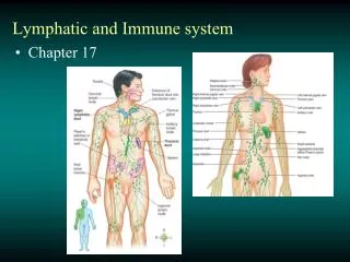
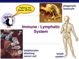
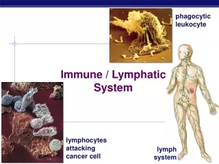
The Lymphatic SystemImmune System • Consists of two semi-independent parts • Lymphatic vessels • Lymphoid tissues and organs • Lymphatic system functions • Transports escaped fluids back to the blood • Plays essential roles in body defense and resistance to disease
Lymphatic Characteristics • Lymph—excess tissue fluid carried by lymphatic vessels • Properties of lymphatic vessels • One way system toward the heart • No pump • Lymph moves toward the heart • Milking action of skeletal muscle • Rhythmic contraction of smooth muscle in vessel walls
Venous system Arterial system Heart Lymph duct Lymph trunk Lymph node Lymphatic system Lymphatic collecting vessels, with valves Lymph capillary Tissue fluid (becomes lymph) Blood capillaries Loose connective tissue around capillaries Figure 12.1
Tissue fluid Tissue cell Lymphatic capillary Blood capillaries Arteriole Venule (a) Figure 12.2a
Lymph • Harmful materials that enter lymph vessels • Bacteria • Viruses • Cancer cells • Cell debris
Lymph Nodes • Filter lymph before it is returned to the blood • Defense cells within lymph nodes • Macrophages—engulf and destroy foreign substances • Lymphocytes—provide immune response to antigens
Entrance of right lymphatic duct into right subclavian vein Regional lymph nodes: Cervical nodes Internal jugular vein Thoracic duct entry into left subclavian vein Axillary nodes Thoracic duct Aorta Spleen Cisterna chyli (receives lymph drainage from digestive organs) Inguinal nodes Lymphatics KEY: Drained by the right lymphatic duct Drained by the thoracic duct Figure 12.3
Tonsils (in pharyngeal region) Thymus (in thorax; most active during youth) Spleen (curves around left side of stomach) Peyer’s patches (in intestine) Appendix Figure 12.5
Spleen • Located on the left side of the abdomen • Filters blood • Destroys worn out blood cells • Forms blood cells in the fetus • Acts as a blood reservoir
Thymus Gland • Located low in the throat, overlying the heart • Functions at peak levels only during childhood • Produces hormones (like thymosin) to program lymphocytes
Tonsils • Small masses of lymphoid tissue around the pharynx • Trap and remove bacteria and other foreign materials • Tonsillitis is caused by congestion with bacteria
Peyer’s Patches • Found in the wall of the small intestine • Resemble tonsils in structure • Capture and destroy bacteria in the intestine
Surface Membrane Barriers:First Line of Defense • Skin and mucous membranes • Physical barrier to foreign materials • Also provide protective secretions • pH of the skin is acidic to inhibit bacterial growth • Sebum is toxic to bacteria • Vaginal secretions are very acidic • Antimicrobial proteins – lysozyme – in saliva, tears • Cilia in trachea • Gastric juice • Symbiotic bacteria in gastric tract
Non-specific barriers • Phagocytes – engulf antigens, neutrophils, monocytes, NK cells (attack tumors) • Complement proteins – lyse the cell wall of the antigen • Interferons – produced by already invaded cells, inhibit viral replication • Inflammatory response – swelling, histamine release, vasodilation
Types of Immune Cells • Immune cells target specific antigens • Lymphocytes – B cells and T cells • B-cells – originate and mature in bone marrow. Produce antibodies • T-cells – produced in bone marrow, reside in thymus • MHC (major histocompatibility complex) markers are on plasma membranes and distinguish self vs. non self cells
Cell-Mediated Response • T cells recognize the antigen MHC markers • They activate other T-cells – memory and helper • The T-helpers activate B-cells – AIDS wipes out T-helpers • T-memory cells “remember” if they have “seen” the virus before • Cytotoxic T-cells kill infected cells
Humoral Response • Antibody mediated response • Immunoglobulins – 5 classes • When B-cells encounted macrophages that are displaying antigen MHC markers • Some B-cells become memory cells for those markers • They produce antibodies that bind to antigens • They will work for future infections