Download
1 / 17
170 likes | 291 Views
This proposal outlines the design and specifications of a cutting-edge microdiffraction instrument for the NSLS X13B beamline, utilizing the Mini-Gap Undulator (MGU) source. The instrument will provide high-resolution focused X-ray beams at micron-scale with both pink beam and monochromatic operation modes. A modular and compact design enables the use of various X-ray optics for flexibility in experiments. High reflecting Kirkpatrick-Baez mirrors and innovative optical components will facilitate low and high divergence radiation, addressing diverse research needs in materials science and biology.

E N D
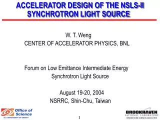
Proposed NSLS X13B Microdiffraction Instrument Source & Optics James M. Ablett National Synchrotron Light Source
X13 Straight-Section EPW MGU
NSLS 'Hard' X-Ray Sources 1st Harmonic ◊MGU ◊ IVUN X17 Wiggler 2nd Harmonic X21 / X25 Wigglers Log [ Brightness ( ph / sec / mm2 /mrad2 / 0.1% BW / 300 mA) ] 3rd Harmonic X13 Wiggler NSLS Bending Magnet Photon Energy [keV]
X13 Undulator Source The Mini-Gap Undulator [MGU] – the brightest source of hard x-rays (E ~ 3.7 keV –16 keV) at the NSLS. MGU parameters: 54 pole, 1.25 cm period At a gap of 3.3 mm , Magnetic field = 0.92 Tesla and Deflection Parameter, K~1.07 Fundamental @ 3.7 keV, 4x10¹⁷ ph/sec/mm2/mrad2/0.1%BW/300mA
MGU Spectral Measurements (August 2002 Ablett,Berman) MGU Brightness – Magnetic Gap=3.3 mm MGU Tuning Curves 18 8x10¹⁷ Calculated 6x10¹⁷ 17 Experiment 16 4x10¹⁷ Brightness (ph/sec/mm2/mrad2/0.1%BW/300mA) Log[Brightness (ph/sec/mm2/mrad2/0.1%BW/300mA)] 15 2x10¹⁷ 14 0 2 4 6 8 10 12 14 16 2 6 10 14 18 Photon Energy [keV] Photon Energy [keV]
Current X13B Beamline Status monochromator hutch 27 24 20 15 12 numbers (in meters )indicate distance from center of X13 straight-section, where MGU is located.
Specifications • The X13B microdiffraction instrument will be modular and compactin design, which will make available a variety of x-ray optics forthe experimentalist. • Focused X-ray Beams on the micron-length scale and below, • together with low and high x-ray divergence will be available with these optics.These will be mounted on a motorized rails and will bereadily accessible. • Pink Beam (Laue) and Monochromatic modes of operationwill be available.- Energy tunability will be accomplished through a 4 bounce monochromator.
X13B Microdiffraction Instrument Layout x-ray microfocusing optics detector circle four-bounce mono tank vibration isolation table diffractometer vibration isolation table 24 25 26 27 Distance from MGU source [meters]
Suite of Optics offered • Pin Holes • Capillaries • KB Mirrors • Zone Plates • Planar Refractive Lenses (Current R&D)
Key Features: • Achromatic - Energy independent focusing-pink beam and monochromatic modes • Accurate Elliptical Figures Achievable (either by bending ormetal deposition ) – Small Spot Sizes • High Reflectivity at Grazing Incidence Angles Kirkpatrick-Baez Elliptical Mirrors Heavy metal Polished spherical Silicon substrate
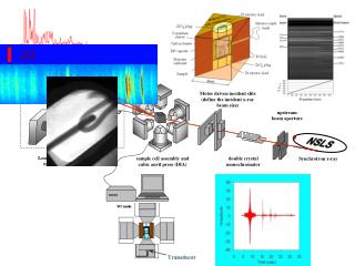
X13B Microprobe …differentially-deposited elliptical KB optics have been used at X13Bduring the past few years, designed primarily for high-pressure x-ray spectroscopy. Vertical blade scan current derivative Horizontal blade scan current derivative ~ 9 microns FWHM ~ 3 microns FWHM blade position [ microns ] blade position [ microns ]
Calcium K Fluorescence Mapping of Osteoarthritic Bone 1. 3.7 mm 3. 1. 2. 2. 3.3 mm 3.
Preliminary KB layout 25 m to MGU source 2cm incident x-rays focal position 3cm 10 cm Expected Parameters 1 micro-radian figure error Spot size – 0.7 x 2 micron2 Divergence – 1mrad (v) x 7 mrad(h) 8 keV, 6x10⁹ ph/sec Gain 5x10⁴ With Figure Error (1 micro-radian)
X13B Microprobe R&D Efforts -Planar Refractive Lenses (K. Evans-Lutterodt, J.M. Ablett) Intensity [arb. Unit] Position [mm]
Summary A state-of-the-art hard x-ray microdiffraction instrumentis proposed for NSLS mini-gap undulator beamline X13B.It will offer a variety of x-ray microfocusing optics –micron scale spatial resolution and below & high and low divergence. Big-beam (unfocused ) mode. Pink Beam and Monochromatic Modes of Operation. Simultaneous fluorescence and diffraction information.