Data Acquisition and Preprocessing in fMRI: A Comprehensive Workshop Overview
This workshop, hosted by Dartmouth College's Department of Psychological and Brain Sciences in collaboration with Yale MR Imaging Center and Kennedy Krieger Institute, focuses on the essentials of fMRI data acquisition and preprocessing techniques. Participants will explore the anatomy of functional data, including slice orientations and localizer sequences, and learn critical preprocessing steps such as motion correction, spatial smoothing, and normalization to align brain images. The workshop also covers statistical analysis, noise reduction, and effective methods to identify brain structures, providing a foundational understanding for effective fMRI research.

Data Acquisition and Preprocessing in fMRI: A Comprehensive Workshop Overview
E N D
Presentation Transcript
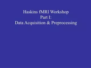
Haskins fMRI WorkshopPart I:Data Acquisition & Preprocessing
Dartmouth College Department of Psychological and Brain Sciences Collaborations Yale University Yale MR Imaging Center Kennedy Krieger Institute F.M. Kirby Research Center for Functional Brain Imaging
Magnet... Source: www.howstuffworks.com Source: http://www.simplyphysics.com/ flying_objects.html
3D space definitions Standard coordinates are listed as mm distances from the origin (the anterior commisure) along the x/y/z dimensions. some examples: Broca’s area, left hemisphere Tal x=-54 y=27 z=9 right cerebellum: Tal x=33 y=-45 z=-39 left occipitotemporal region: Tal x=-39 y=-45 z=-19 • http://www.sph.sc.edu/comd/rorden/anatomy/home.html
slice orientations sagittal coronal axial
Typical Acquisition Sequence three-plane “localizer” sagittal “scout” axial T1 anatomic several functional runs... high-resolution anatomic (MP-RAGE) Diffusion Tensor Imaging....
Simulated Hemodynamic Response Noise SD = 0 Noise SD = 10 Noise SD = 100
Preprocessing steps functional data: • adjust for slice acquisition time sinc interpolation; “temporal realignment” • adjust for motion “motion correction”; “(spatial) realignment” • apply spatial smoothing gaussian filter anatomic data: • strip skull from image • align with a common template “normalization”
slice acquisition time... We typically acquire 20 (functional) slices in each 2-second interval. Each one takes 100 msec. Must account for this in analysis... functional data: • adjust for slice acquisition time acquisition order 2 1 slice #1 (circles) is acquired at times 0/2/4/6/8... seconds exactly. slice #2 (diamonds) is acquired 100msec later, at times 0.1/2.1/4.1/6.1/8.1... seconds post-stimulus. stimulus onset at time 0
normalization Basic idea: find a transformation that will spatially shift this subject’s brain to align with a template, so that subjects can be averaged together. This also allows us to use a pre-labelled atlas to identify structures. important concepts: • spatial transformations: linear: translation, rotation, scaling; nonlinear: warps • degrees of freedom (DOF) 6: Rigid Body; 7: adds global rescale; 12: affine (adds shear) • similarity functions; search & optimization; resolutions • skull stripping