
The Integumentary System
The Integumentary System. Objectives. To identify the components of the integumentary system To identify layers of skin and their function To identify and determine the function of the accessory structures To identify common disorders and treatments of the integumentary system.

The Integumentary System
E N D
Presentation Transcript
Objectives • To identify the components of the integumentary system • To identify layers of skin and their function • To identify and determine the function of the accessory structures • To identify common disorders and treatments of the integumentary system
What is the Integumentary system? • Derives its name from the Latin word “integumentum,” which means “to cover” • Is the largest, heaviest organ in the body • Is absolutely necessary for life This is what your body would look like without the integumentary system!
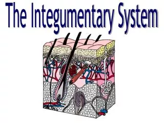
What makes up the Integumentary System? • Skin • epidermis • dermis • hypodermis • Accessory Structures • hair • nails • exocrine glands
Skin = Integument = Cutaneous Membrane 7 Functions: • Protective covering • Regulates body temperature • Manufactures Vitamin D • Sensory function • Temporary storage of fat, glucose, water and salts • Screens out harmful ultraviolet radiation • Absorbs certain drugs
2 basic layers Epidermis Outermost covering Epithelial cells Avascular Produced vitamin D Dermis True skin Connective tissue Vascular STRUCTURE OF THE SKIN
Hey— your epidermis is showing!!
Epidermis Take a look at your hands. What do you see happening? Although you are not able to see it, you epidermis is constantly working around the clock. The bottom layer of the epidermis is hard at work synthesizing new skin cells. When the new cells are developed, they begin making their way toward the outer layer of the epidermis. A continuous pattern develops. New cells are constantly made and help push the dead cells upward. As old cells die near the top, they rise to the surface of your skin. So, what you really see when you look at your skin are dead skin cells.
EPIDERMIS2 (of 3) epidermal layers are:Stratum corneumStratum germinativum STRATUM CORNEUM • Outermost layer • In cells, cytoplasm replaced by KERATIN – making them waterproof. • Flat and scale-like cells that flake off • First line of defense against surface bacteria • Thickest on palms of hands, soles of feet STRATUM GERMINATIVUM • Innermost layer • Reproductive layer – cells form and push their way up, become keratinized, and replace the top layer • Contains MELANOCYTES – cells that contain a pigment = MELANIN
Melanin • Black, brown, or has a yellow tint – depending on racial origin • Protects the body against UV radiation • Absorbs UV light before damage occurs to cells’ DNA The more melanin, the darker the skin Caucasians don’t have much melanin in their melanocytes. Freckles = patches of melanin Albinism = no melanin
PAPILLAE • Ridges in stratum germinativum that arise from dermis • Create permanent ridges in fingers, palms and soles of feet • These “friction ridges” help with grip • Cause “fingerprints”
DERMIS • Is the second thicker layer of skin • Contains the accessory structures • Hair • Sweat glands • Oil glands • Contains blood vessels, which supply nutrients and oxygen to the cells • Contains sensory receptors (nerves) for: • touch • pressure • hot and cold • pain
Nerve Receptors in Dermis Sensory nerves – heat, cold, touch, pain and pressure Touch receptors - close to the surface Pressure receptors - are deeper
Distributed over the entire skin surface Large numbers under the arms, palms of hands, soles of feet and forehead Duct extends to form a pore in the skin, perspiration excreted through the pores May be activated by heat, pain, fever and nervousness Average fluid loss is 500 ml per day Perspiration is 99% water Sweat Glands
Sebaceous Glands • Produce a mixture of lipids and proteins called sebum • Function to prevent drying of skin and hair • Are located primarily on the scalp, face and upper portion of the body • Increase in size and produce a greater amount of sebum in coordination with increased hormone levels
Hypodermis • Is also called the subcutaneous layer • Is the innermost layer of skin • Is composed primarily of fat • Acts as a shock absorber • Anchors skin to underlying muscle
Hypodermis • Origin of hair follicles • follicles are tiny tubes in which body hair grows out of • follicles are found all over your body, except the lips, palms of the hands and soles of the feet
So why do my fingers wrinkle when I take a bath? Your skin does have a waterproof coating of sebum and sweat. However, when you are exposed to water for long periods of time, this waterproof coating washes away. Once this coating is washed away, your skin can absorb water. When your skin absorbs water it gets soggy and wrinkly. After you dry off, your skin will begin to replenish the layer of sebum and sweat.
HAIR Fibers, non-living protein structures Amount & type are species and environment dependant Has capillaries and nerves attached to follicle, which provide nutrients and sensory information Outer layer = CORTEX Inner layer = MEDULLA Part under the skin = ROOT Part outside the skin = SHAFT FOLLICLE = “pocket” in epidermis, hair inside PAPILLA = tuft of tissue in root, contains capillaries ARRECTOR PILI MUSCLE = smooth muscle attached to follicle. Appendages of the Skin
Q: How does this muscle cause goose bumps? A: When they contract, they pull the hair into an upright position, causing skin dimples (goose bumps). The nervous system regulates these muscles; cold temperatures or fright can activate them.
What makes my hair shiny? At the dermis layer, each follicle has its own sebaceous gland. The sebaceous gland releases sebum onto the hair. Sebum lightly coats your hair, making it shiny and slightly waterproof.
Nail • Develop from the epidermis • Formed in the nail bed or MATRIX • Epidermal cells fused together and fill with keratin • Appear pink because their translucency reveals the vascular tissue beneath. • They aid in grasping, scratching, and protecting fingers and toes.
Components: The lunula is the white half-moon at the nail base. The body and free edge overhangs the end The nail rests on the thick layer of epithelial skin called the nail bed. The hyponychium is the epithelium of the nail bed The root is hidden under skin Under the root lies the matrix (thick layer of skin). Eponychium (thin layer of epithelium) covers the nail during development; in the adult, it remains at the nail base only and is called the cuticle.
Skin and Microorganisms • Intact skin = best protection against pathogens, toxins and water loss • Skin generally too dry for microbial growth – they do grow in moist areas • Most skin bacteria associated with hair follicles or sweat glands • Underarm perspiration odor caused by bacteria and perspiration The best way to prevent the spread of disease is by hand washing.
Temperature Regulation • If the body is too hot and needs to release heat: • A signal from the hypothalamus (part of the brain) is sent to the skin and warm blood is brought closer to the surface of the skin • Sweat glands begin producing lots of sweat to release into the air, cooling the body • When the body is cold and needs to conserve heat: • The brain senses cold body temperatures • Blood vessels constrict to keep warm blood away from the surface of skin • Pilomotor reflex occurs– formation of “goose bumps”
Fun Facts About Your Skin • An adult’s skin weighs about 5-10 pounds • If your skin was spread out like a blanket, it would measure about 20 square feet • Each minute we loose 30,000 – 40,000 skin cells off the surface of our skin • There are more than 100,000 hair follicles on your head
ACNE ATHLETE’S FOOT DERMATITIS ECZEMA WARTS (VERRUCAE) GENITAL HERPES IMPETIGO PSORIASIS RINGWORM URTICARIA / HIVES BOILS (CARBUNCLES) SHINGLES (Herpes Zoster) HERPES SIMPLEX 1 GENITAL HERPES SCABIES SKIN CANCER BASAL & SQUAMOUS CELL CARCINOMA, MALIGNANT MELANOMA Disorders of the Skin
ACNE • Disorder of the sebaceous glands • Sebum plugs pores and area fills with leukocytes • Blackheads, cysts, pimples, and scarring
Boils • a.k.a. Carbuncles • Painful, bacterial infection
Athelete’s Foot • Contagious fungal infectious • Usually contracted in public baths and showers • Rx – antifungal agents
Dermatitis • Non-specific skin inflammation • Rash – reaction to soap, plants, etc. • Skin blotches – caused by stress
Eczema • Acute or chronic inflammatory skin disease • Skin dry, red, itchy and scaly • Rx – remove cause, hydrocortisone to help alleviate symptoms
Herpes • Genital herpes • Viral blister in genital area • Spread through sexual contact • Periods of remission & exacerbation • Rx: Acyclovir • Can be passed to newborn during delivery • Herpes Simplex I • Virus • Fever blister or coldsore • Shingles (zoster) • Viral infection of nerve endings • On chest or abdomen, accompanied by severe pain
Impetigo • Acute, inflammation and contagious • Seen in babies and young children • Caused by staphylococcus or streptococcus • Vesicles that rupture and develop distinct yellow crusts
Psoriasis • Chronic inflammatory skin disease • Dry reddish patches covered with silvery-white scales
Ringworm • Contagious fungal infection • Raised, itchy, circular patches with crusts
Scabies • Communicable • Severe itching • Mite burrows in skin, lays eggs, eggs hatch
Tanning • Sunlight stimulates melanocytes to make more melanin • Tanning produced by UV rays. • Prolonged exposure may lead to skin cancer!
SKIN CANCERAssociated with exposure to sun (UV rays)***Most common type of cancer in people*** • BASAL CELL CARCINOMA • Most common, least malignant • Usually on face • Rx: surgery or radiation • SQUAMOUS CELL CARCINOMA • Mostly scalp and lower lip • Grows rapidly and metastasizes to lymph nodes • Rx: surgery or radiation • Prognosis good with early diagnosis
SKIN CANCER (cont) • MALIGNANT MELANOMA • Occurs in melanocytes • Metastasizes to other areas quickly • Appears as brown or black irregular patch that occurs suddenly • A change in an existing wart or mole • RX: surgical removal of melanoma and surrounding area & chemotherapy
Skin Lesions • Pustule – acne • Ulcer – venous stasis ulcer superficial or decubitus • Tumor – benign epidermal tumor, basal cell carcinoma • Vesicle – chicken pox, herpes simplex Other Terms: Excoriation – abrasion Pruritis - Itching
First Degree • Superficial • Skin red and dry • Involves only epidermis • Rx – cold water • Healing within one week
Second Degree • Epidermis and dermis • Pain, swelling, redness and blistering • Skin may be exposed to infection • Rx – pain medication, dry sterile dressing • Healing within 2 weeks
Third Degree • Epidermis, dermis and subcutaneous layers • Symptoms – loss of skin, blackened skin • May be life threatening
RULE OF NINESMeasures percent of body burned. Body divided into 11 area, each is 9% of body surface.


