Download
1 / 30
300 likes | 436 Views
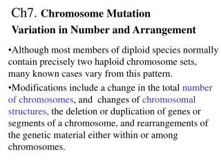
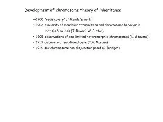
Development of chromosome theory of inheritance ~1900 “rediscovery” of Mendel’s work 1902 similarity of mendelian transmission and chromosome behavior in mitosis & meiosis (T. Boveri; W. Sutton) 1905 observations of sex-limited heteromorphic chromosomes (N. Stevens)

E N D
Development of chromosome theory of inheritance • ~1900 “rediscovery” of Mendel’s work • 1902 similarity of mendelian transmission and chromosome behavior in • mitosis & meiosis (T. Boveri; W. Sutton) • 1905 observations of sex-limited heteromorphic chromosomes (N. Stevens) • 1910 discovery of sex-linked gene (T.H. Morgan) • 1916 sex chromosome non-disjunction proof (C. Bridges)
Development of chromosome theory of inheritance • ~1900 “rediscovery” of Mendel’s work • 1902 similarity of mendelian transmission and chromosome behavior in • mitosis & meiosis (T. Boveri; W. Sutton) • 1905 observations of sex-limited heteromorphic chromosomes (N. Stevens) • 1910 discovery of sex-linked gene (T.H. Morgan) • 1916 sex chromosome non-disjunction proof (C. Bridges) replication replication meiosis mitosis Fig. 3-1
Drosophila melanogaster has a heteromorphic chromosome pair • Males have one of each member (XY) • Females have two of one member of the pair (XX) Fig. 3-5
Morgan’s white gene analysis white-eye male X red-eye female F1 all red-eye F2 ½ males red-eye ½ males white-eye all females red-eye red = w+ allele (wildtype) white = w allele (mutant)
Morgan’s white gene analysis Reciprocal cross white-eye male X red-eye female F1 all red-eye F2 ½ males red-eye ½ males white-eye all females red-eye red-eye male X white-eye female F1 all males white-eye all females red-eye F2 ½ males red-eye ½ males white-eye ½ females red-eye ½ females white-eye Sex-dependent transmission & expression (sex-linkage)
Morgan’s white gene analysis - INTERPRETATION Reciprocal cross white-eye male X red-eye female Xw/Y Xw+/Xw+ F1 all red-eye Xw+/Y Xw/Xw+ F2 ½ males red-eye Xw+/Y ½ males white-eye Xw/Y all females red-eye Xw+/Xw Xw+/Xw+ red-eye male X white-eye female Xw+/Y Xw/Xw F1 all males white-eye Xw/Y all females red-eye Xw/Xw+ F2 ½ males red-eye Xw+/Y ½ males white-eye Xw/Y ½ females red-eye Xw+/Xw ½ females white-eye Xw/Xw Sex-linkage indicates locus on a sex-limited chromosome
Sex linkage can be the reverse (e.g., lepidopterans, birds) • Females have the heteromorphic chromosome pair (ZW) • Are the heterogametic sex • Males are ZZ • Are the homogametic sex Fig. 3-6
C. Bridges’ cross of red males (w+/Y) and white females (w/w) produced rare • white female (must be w/w = gets two X chromosomes from mother?) • red male (must be w+) = gets father’s X chromosome?
C. Bridges’ cross of red males (w+/Y) and white females (w/w) produced rare • white female (must be w/w = gets two X chromosomes from mother?) • red male (must be w+) = gets father’s X chromosome? Explanation: chromosome nondisjunction Fig. 3-7 • Confirmed chromosome theory • Discovered general class of rare, recurring mutation with significant consequences • Demonstrated why geneticists should “treasure your exceptions”
The sizes and densities of genes differs considerably among organisms Fig. 3-13 • Due to: spacing between genes • introns, relative number and sizes • other stuff
Genic organization of two small regions of human chromosome 21 Fig. 3-12
Karyotype of Indian muntjac (2n+6) Fig. 3-8
Conventional cytogenetic markers for karyotype analysis • Chromosome number (n, 2n, etc.)
Conventional cytogenetic markers for karyotype analysis • Chromosome number (n, 2n, etc.) • Chromosome size (large → small) • Centromere location: central (metacentric) • terminal (telocentric) • in-between (acrocentric) • Chromatin differential compaction • Euchromatin (lightly staining, lower density of DNA, most genes) • Heterochromatin (densely staining, highly compacted,usually centromeric) • Banding patterns (localized, alternating high/low compaction regions)
Drosophila karyotype in diploid and polytene cells (larval salivary gland cells) Fig. 3-18
Conventional cytogenetic markers for karyotype analysis • Chromosome number (n, 2n, etc.) • Chromosome size (large → small) • Centromere location: central (metacentric) • terminal (telocentric) • in-between (acrocentric) • Chromatin differential compaction • Euchromatin (lightly staining, lower density of DNA, most genes) • Heterochromatin (densely staining, highly compacted,usually centromeric) • Banding patterns (localized, alternating high/low compaction regions) • Special structures (nucleolus organizers, constrictions) Stylized corn chromosomes Fig. 3-19
In situ hybridization with a fluorescently labelled probe for a single-copy muscle protein gene Fig. 3-1 Fig. 3-11
In situ hybridization with a 3H-labelled probe for a mouse satellite DNA Fig. 3-14
In situ hybridization with a fluorescently labelled telomeric DNA sequence (human fibroblast) Fig. 3-16
Repeated from Cell Biology (must read/review) Pages 88-90: chromosome/chromatin structure Pages 90-95: mitosis & meiosis Pages 96-97: life cycles of haploid and diploid organisms
Overview of allele transmission through mitosis and meiosis Fig. 3-35
Overview of allele transmission through chromatid formation/ DNA replication Fig. 3-36
Euglena genomes • nuclear (red) • mitochondrial (yellow) Fig. 3-40
Neurospora nuclear (ad) and mitochondrial (poky) genes display distinctive sexual transmission patterns (maternal inheritance) Fig. 3-42
Organellar genes can show cytoplasmic segregation during asexual growth Fig. 3-43
Typical inheritance of a human mitochondrial disease Fig. 3-45