Protein Folding and Processing
Protein Folding and Processing. The classic principle of protein folding is that all the information required for a protein to adopt the correct three-dimensional conformation is provided by its amino acid sequence.

Protein Folding and Processing
E N D
Presentation Transcript
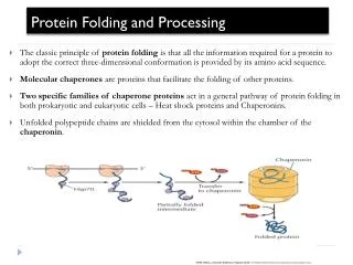
Protein Folding and Processing • The classic principle of protein folding is that all the information required for a protein to adopt the correct three-dimensional conformation is provided by its amino acid sequence. • Molecular chaperones are proteins that facilitate the folding of other proteins. • Two specific families of chaperone proteins act in a general pathway of protein folding in both prokaryotic and eukaryotic cells – Heat shock proteins and Chaperonins. • Unfolded polypeptide chains are shielded from the cytosol within the chamber of the chaperonin.
Action of chaperones during translation and Transport • chains that are still being translated on ribosomes, thereby preventing incorrect folding or aggregation of the amino-terminal portion of the polypeptide before synthesis of the chain is finished. • Chaperones also stabilize unfolded polypeptide chains during their transport into subcellular organelles.
Protein folding in the cell Basics - cell compartments, molecular crowding: cytosol, ER, etc. Folding on the ribosome - co-translational protein folding Molecular chaperones - concepts, introduction - intramolecular chaperones - chemical chaperones - protein chaperones
Folding in vitro vs.in vivo in vitro in vivo protein denatured in a chaotrope folding by dilution in buffer folding folded protein folded protein
Problem: non-native proteins exposed hydrophobic residues X X X X incorrect molecular interactions & loss of activity intramolecular intermolecular X X X X X X misfolding aggregation 3-10 • non-native proteins expose hydrophobic residues that are normally buried within the ‘core’ of the protein • these hydrophobic amino acids have a strong tendency to interact with other hydrophobic (apolar) residues - especially under crowding conditions
The Unfolded Protein Response (UPR) Hsp4 (grp78) grp170 XBP-1 IRE-1 • The UPR occurs when proteins are misfolded in the endoplasmic reticulum (ER). • Reducing agents, such as DTT, interfere with disulfide bond formation while drugs can interfere with glycosylation; both agents cause proteins to misfold in the ER thus triggering the UPR. • The product of the ire-1 gene is the sensor of misfolded proteins and when activated removes an intron from the pre mRNA from the xbp-1 gene. • Active xbp-1 protein (from spliced mRNA) activates the genes that code for ER chaperones, such as hsp-4.
Degradation of proteins • 1) dietary proteins • - amino acids • - pepsin in stomach • - pancreatic proteases • - aminopeptidase N • other peptidases • 2) endogenous proteins • - protein turnover: synthesis, degradation, resynthesis • - damaged proteins • - half-lives of proteins: depend on amino-terminal residues PROTEIN TURNOVER AND AMINO ACID CATABOLISM
Cellular Protein Degradation • Lysosomal • Nonspecific • Endocytosis • Foreign proteins • Energy favorable to degrade proteins • Non-lysosomal • Specificity, requires ATP • Conditions of stress • Ubiquitin-proteosomal pathway • 26S proteosome • Role in cellular processes/signaling
Protein turnover; selective degradation/cleavage Individual cellular proteins turn over (are degraded and re-synthesized) at different rates. E.g., half-lives of selected enzymes of rat liver cells range from 0.2 to 150 hours. N-end rule: On average, a protein's half-life correlates with its N-terminal residue. • Proteins with N-terminal Met, Ser, Ala, Thr, Val, or Gly have half lives greater than 20 hours. • Proteins with N-terminal Phe, Leu, Asp, Lys, or Arg have half lives of 3 min or less. PEST proteins having domains rich in Pro (P), Glu (E), Ser (S), Thr (T), are more rapidly degraded than other proteins.
Ubiquitinylation – Proteosome Degradation E3 determines protein substrate
Ubiquitination • 1) ubiquitin • - a 8.5 kd protein (76 residues) • formation of an isopeptide bond with ε-amino group of lysine • of the proteins • - a tag for destruction • - polyubiquitin: a strong signal for degradation • 2) enzymes for ubiquitination • - E1 (ubiquitin-activating enzyme) • - E2 (ubiquitin-conjugating enzyme) • - E3 (ubiquitin-protein ligase) • - variation: E3 > E2 > E1: more finely tuned substrate discrimination • HPV (human papilloma virus) activates a specific E3 enzyme: • tumor suppressor protein p53
Regulation of ubiquitination: Some proteins regulate or facilitate ubiquitin conjugation. Regulation by phosphorylation of some target proteins has been observed. E.g., phosphorylation of PEST domains activates ubiquitination of proteins rich in the PEST amino acids. Glycosylation of some PEST proteins with GlcNAc has the opposite effect, prolonging half-life of these proteins.
19S and 20S Proteasome Subunits Characteristics • 20S Subunit • Barrel • Contains 6 proteolytic sites • 2x Tryptic • 2x Chymotryptic • 2x Peptidylglutamyl- peptidase • Linearized protein required • 19S Subunit • Base and Lid • Contains subunits with known and unknown functions • Tetra-Ub (K48) recognition • Deubiquitination activity • Protein unfolding activity (Chaperone function)
Ubiquitin AA Sequence MQIFVKTLTG KTITLEVEPS DTIENVKAKI QDKEGIPPDQ QRLIFAGKQL EDGRTLSDYN IQKESTLHLV LRLRGG 6 48 63
Proteasome-1 Proteasome-3 Proteasome-4
Transmembrane Proteins Regulated by Ub-dependent Sorting In metazoans: Neurotransmission:Ion channels: AMPA glutamate receptors ENaC Glycine receptors ClC-5 Cell-cell contacts:Immune molecules E-cadherin downregulated by viruses: Occludin MHC class I B7-2 Developmental patterning: ICAM-1 Delta CD4 Notch Roundabout
Ub Ub Ub Ub Poly-Ub Chains K48 Linkage K63 K Signal to proteosome K48, Ub4 K63 Linkage K48 K Ub Ub Cell Signaling K63 Ub Peters, J.M. 1998 Ubiquitin and the Biology of the Cell Ub Ub
ENaC function • Major ion channel that controls salt and fluid resorption in the kidney • Mutations in the PPXY motif cause accumulations of channels at the cell surface and result in Liddle’s syndrome, and inherited form of hypertension
ENac surface Stability • Nedd 4 (HECT ligase)-negatively regulates ENaC surface stability • Nedd4 WW domains bind PPXY motif of ENaC subunits • Nedd4 also interacts with serum and glucocorticoid-regulated kinase (SGK) • SGK contains two PPXY motifs that bind to Nedd4 WW domains • SGK-dependent Nedd4 P inhibits the Nedd4-ENaC interaction • therefore, Nedd4 P increases ENaC at the cell surface
Ub-like Proteins • SUMO-1 (sentrin, smt-3) • 1996 – covalent modification – RanGAP1 • RanGAP1 nearly quantitative modified • Cytosolic RanGAP1 to nuclear pore • Activate shuttling factor
Ubiquitin Superfold and Ubiquitons UB αβ roll suprfold Ub – blue SUMO-1 – green NEDD8 - red
SUMO • SUMO • SUMO-1 & SUMO-2/3 • Shared characteristics • C-terminal -GG essential for conjugation • Affix to lysine residues in target • NOT directly associated with proteasomal degradation
Competition/Regulation SUMO Reactive Oxygen Species: Oxidizes reactive thiols on SUMO enzymes Uba1/Aos1- S – S – Ubc9 Thus: SUMO can not attach and proteins not Sumoylated
Examples of SUMO function • RanGAP • IkB • c-Jun • p53 and mdm2 • Causes nuclear translocation • Blocks Ub-conjugation site, prevents degradation • Inhibits transcriptional activity • Blocks mdm2 self-ubiquitination, prevents degradation • SUMO-p53 in DNA binding domain apoptotic activity SUMO Effect PROTEIN
Proteasome specificity • NetChop is the best available cleavage method • www.cbs.dtu.dk/services/NetChop-3.0