Download
1 / 29
300 likes | 509 Views
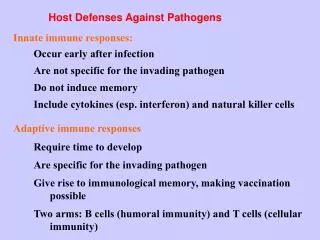
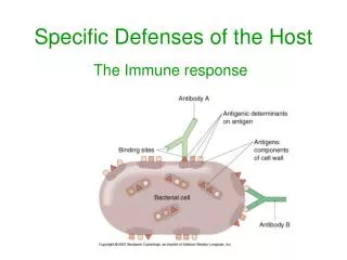
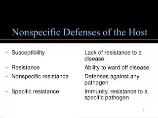
Nonspecific Defenses of the Host. Susceptibility: Lack of resistance to a disease. Immunity: Ability to ward off disease. Innate immunity: Defenses against any pathogen. Adaptive immunity: Immunity, resistance to a specific pathogen. Host Defenses. Figure 16.1. Physical Factors. Skin

E N D
Nonspecific Defenses of the Host • Susceptibility: Lack of resistance to a disease. • Immunity: Ability to ward off disease. • Innate immunity: Defenses against any pathogen. • Adaptive immunity: Immunity, resistance to a specific pathogen.
Host Defenses Figure 16.1
Physical Factors • Skin • Epidermis consists of tightly packed cells with keratin, a protective protein
Physical Factors • Mucous membranes • Ciliary escalator: Microbes trapped in mucus are transported away from the lungs. • Lacrimal apparatus: Washes eye. • Saliva: Washes microbes off. • Urine: Flows out. • Vaginal secretions: Flow out. Figure 16.4a
Chemical Factors • Fungistatic fatty acid in sebum. • Low pH (3-5) of skin. • Lysozyme in perspiration, tears, saliva, and tissue fluids—what does it do? • Low pH (1.2-3.0) of gastric juice. • Transferrins in blood find iron. • NO inhibits ATP production.
Normal Microbiota • Microbial antagonism/competitive exclusion: Normal microbiota compete with pathogens.
Formed Elements in Blood Table 16.1 (1 of 2)
Formed Elements in Blood Table 16.1 (2 of 2)
Differential White Cell Count • Percentage of each type of white cell in a sample of 100 white blood cells.
White Blood Cells • Neutrophils: Phagocytic • Basophils: Produce histamine • Eosinophils: Toxic to parasites (worms), phagocytosis • Dendritic cells: Initiate adaptive immune response • Monocytes: Phagocytic as mature macrophages • Fixed macrophages in lungs, liver, and bronchi • Wandering macrophages roam tissues. • Lymphocytes: Involved in specific immunity.
Phagocytosis • Phago: from Greek, meaning eat • Cyte: from Greek, meaning cell • Ingestion of microbes or particles by a cell, performed by phagocytes. Figure 16.6
Phagocytosis Figure 16.7
Inflammation • FOUR CARDINAL SIGNS: • Redness • Pain • Heat • Swelling (edema) • Acute-phase proteins activated (complement, cytokine, and kinins) • Vasodilation (histamine, kinins, prostaglandins, and leukotrienes) • Margination and emigration of WBCs • Tissue repair
Inflammation Figure 16.8a–b
Inflammation Figure 16.8c–d
Fever: Abnormally High Body Temperature • Hypothalamus normally set at 37°C. • Gram-negative endotoxin cause phagocytes to release interleukin–1 (IL–1). • Hypothalamus releases prostaglandins that reset the hypothalamus to a high temperature. • Body increases rate of metabolism and shivering which raise temperature. • When IL–1 is eliminated, body temperature falls (crisis).
Advantages Increase transferrins Increase IL–1 activity Disadvantages Tachycardia Acidosis Dehydration Fever
The Complement System • Serum proteins activated in a cascade. Figure 16.9
Effects of Complement Activation • Opsonization or immune adherence: Enhanced phagocytosis. • Membrane attack complex: Cytolysis. • Attract phagocytes. Figure 16.10
Effects of Complement Activation Figure 16.11
Classical Pathway Figure 16.12
Alternative Pathway Figure 16.13
Lectin Pathway Figure 16.14
Some Bacteria Evade Complement • Capsules prevent C activation. • Surface lipid-carbohydrates prevent MAC formation. • Enzymatic digestion of C5a.
Interferons (IFNs) • Alpha IFN and Beta IFN: Cause cells to produce antiviral proteins that inhibit viral replication. • Gamma IFN: Causes neutrophils and macrophages to phagocytize bacteria.
Interferons (IFNs) Figure 16.15
Transferrins Bind serum iron Antimicrobial peptides Lyse bacterial cells Innate Immunity