REAL-TIME PCR
350 likes | 691 Views
REAL-TIME PCR. PCR. PCR is a widely used technique for detecting and quantifying DNA from organisms and has become an essential tool in research laboratories and diagnostic tool used in clinical research

REAL-TIME PCR
E N D
Presentation Transcript
PCR • PCR is a widely used technique for detecting and quantifying DNA from organisms and has become an essential tool in research laboratories and diagnostic tool used in clinical research • However, the combination of existing detection assays for PCR suffer from long and tedious post PCR processes • Gel electrophoresis • Hybridization • Blocking, Washing and Rinsing • Cross contamination by subsequent steps • This led to kinetic studies of reactions to devise the real time PCR system
REAL-TIME PCR • A revolutionary method of specific DNA/RNA sequence identification and amplification that can simultaneously quantify the amount of each starting sample • Relies on the detection of specific sequence primers and quantization by fluorescent reporter probes in which the signal increases directly proportional to the amount of PCR product in a reaction • Records the amount of fluorescence emission at each cycle using a computer algorithmic program
APPLICATIONS • Quantization of gene expression • Viral quantization • Pathogen detection • Genotyping • Prenatal diagnosis • Allelic discrimination
ADVANTAGES • Maximizes power of traditional PCR applications • Eliminates post PCR processes • Low turn–around time • Cost effectiveness/practicality of supplies • Minimizes cross–contamination • High sensitivity • needs < 5 copies of DNA/RNA • Dynamic range of quantification • multiplexing • High throughput capacity • A lot of amplifications within a small time frame as opposed to other microbiological methods • Cell culturing • Viral growth
REAGENTS • Traditional PCR reagents • RNA/DNA sample • dH20 • DNA Polymerase • PCR buffer • Mg2+ • Salt • Tris-HCl base at specific pH • dNTPs • Forward and Reverse Primers • Additional real time PCR reagents • Fluorophore labeled probes • Internal control elements
PROBES • The probe is an oligonucleotide that has both a reporter fluorescent dye and a quencher dye attached and is sequence specific to the amplified target • As long as the quencher is in close proximity of the reporter dye the fluorescence emitted by the reporter is blocked • The distance separation of the reporter dye from the quencher allows the fluorescence emission to be emitted and detected by the real time PCR instrument
EXAMPLE OF FLUROPROBES • Taqman [Figure A] • Depend on the 5’ nuclease activity of DNA polymerase for PCR to degrade the hybridized fluroprobe, removing the reporter from the quencher • Molecular Beacon [Figure B] • adopts hairpin loop structure while in solution to bring the reporter dye and quencher dye in close proximity for FRET to occur • once the beacon hybridizes to the target sequence during the annealing step, the reporter dye is separated from the quencher dye and the reporter fluorescents
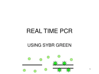
EXAMPLE OF FLUROPROBES • Scorpions • maintains stem–loop configuration when not hybridized • when the stem–loop is opened due to complementary binding of 3’, end a fluorescent signal is observed • SYBR Green • Binds dsDNA, indiscriminate of sequence specificity, and emits signal
INSTRUMENTATION • Thermocycler with optics for fluorescent excitation and emission collection • Computer connected to thermocycler instrument • Analysis software for data acquisition and calculation
DATA ACQUISITION • The computer measures the intensity of each well, individually, by using a sensitive camera to monitor the fluorescence at cyclic intervals during the PCR reaction in the thermocycler
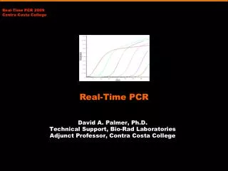
DATA INTERPRETATION • Intensity (Rn) is the measure of fluorescence • this value indicates magnitude of the signal generated • Threshold is the average standard deviation of Rn for the early cycles (background) • Intersection between the amplification plot and the baseline is the cycle threshold or CT value • CT value is the cycle at which a significant increase in the change of Rn is first detected • It is at this cycle, that we can start to compare and analyze the samples relative to each other
DATA INTERPRETATION • A plateau phase represents the point at which DNA is no longer being amplified • It at this cycle that we must stop comparing and analyzing the samples relative to each other. • Exponential phase provides the most useful information about the reaction because it is here that the log-linear phase can be conferred [Figure 32B] • The slope of a log-linear phase (transcribe exponential plot into log-linear) is a direct reflection of the amplification efficiency
CRITICAL PARAMETERS • Specificity • Primers • Must flank the region of amplification • Must not be complementary • Avoid primer dimers • Probe • Specific to a target sequence within the amplified region or to dsDNA • Must carry a quencher and fluorolabeled probe • Sensitivity • Amplification of small amount of sample • Contamination free/Quality control procedure • Adequate PCR environment and reagents • Signal Amplification • For multiplexing dyes – insurance of distinguishing fluorescence emissions • Removal of other agents that give off similar fluorescence readings • Controls • positive control – controls for failure of PCR reagents • negative control – controls for presence of contaminant DNA • Efficiency of Curves • Threshold • Log-linear slopes
IDI-Strep B Real time PCR DNA-based diagnostic test for detecting Group B streptococcus
INTRODUCTION • IDI-Strep B is an assay that can rapidly detect Group B streptococcus (GBS) in pregnant women • Qualitative • Quantitative • Specific through DNA complementary binding • Sensitive (ideally needing only a single copy of DNA) • in vitro • Molecular diagnostic utility detecting DNA (as opposed to culture based testing)
GBS • Group B Streptococcus (GBS) has been recognized as the primary cause of bacterial infection in newborn babies, resulting in life threatening conditions within newborns. • 35-40% of women will carry the GBS bacteria in their urinary tract, digestive tract, and/or urinary tract. • During pregnancy and after delivery, GBS can cause serious illness in both mothers and newborns. • 15% to 20% of pregnant women have a different colonization status at delivery time than at 35-37 weeks • Due to the underdeveloped immune system, primarily in preterm births, GBS poses a leading cause of death within newborns • Life-threatening conditions, such as pneumonia and meningitis, as a few examples, are a result of GBS infections that occur early on in life of newborns (developing within a week) • About half of all infants born with GBS will die and the rest may develop in serious brain damage • 10% term babies and 40% preterm babies • Most cases of GBS infection in newborns can be prevented by giving certain pregnant women antibiotics during labor. • Antibiotic treatment before labor does not prevent GBS infection in newborns.
GBS • How is GBS infection diagnosed? • The current ‘gold standard’ screening test is a rectovaginal culture in selective medium, taken at 35-37 weeks (8.5 months) • Results are available within 2 days using culturing methods in selective medium • Problems: cannot accurately predict genital tract colonization at time of labor. • Risk factor method for diagnosing GBS targets treatments indiscriminately to women believed to be at great risk. • Problem: This is a guess! We would miss many colonized mothers and at risk infants. • Problem: We would economically and production-wise WASTE by giving medication unnecessarily!
GBS Detection • IDI-Strep B – real time PCR • Specifically designed diagnostic test to detect GBS in pregnant women in less than 1 hour of delivery! • Combination of diagnostic benefits • Speed: Less than 1 hour! • Sensitivity: 94% • Specificity: 96% • Simplicity • Uses normal vaginorectal swab techniques • Easy 3 step nucleic acid extraction • Simple real time PCR automated processing
IDI-Strep B • IDI-Strep B uses real time PCR for amplification of the specific cfb gene of GBS coupled simultaneously with fluorogenic target-specific hybridization for detection of amplified DNA • Test/Assay utilizes: • Polymerase chain reaction (PCR) Fluorogenic target-specific hybridization • Computer analysis programs to qualify, validate, and quantify results • Power of the Assay: • Highly specific and sensitive • Increases quantitative power of PCR by measuring fluorescence activity per cycle of amplification • Speed and efficiency of the results • Removal of post-PCR processes • Gel Electrophoresis • X-ray film
IDI-Strep B • Technical background • IDI-Strep B uses the molecular beacon method for probe/fluorescence targeting • 1. Cell Extraction of DNA • A vaginal/rectal specimen is collected • Swab is placed in a sample preparation buffer to elute the contents • An aliquot of the specimen is then lysed and added, as the sample, to the PCR reagents • Entire process takes 15 minutes! Whew, that’s fast!
IDI-Strep B • 2. Primers: Amplification of the GBS gene, cfb, using two GBS-specific primers. • Cfb is a 154bp region found only within GBS! • An internal control (IC) is also used to confirm the integrity of the assay reagents • Similar to the purpose of amplifying beta-globin gene we used in 14;18 translocation
IDI-Strep B • 3. Fluorescence by Hybridization probes • Amplified targets (cfb and IC) are detected with fluorescence labeled hybridization probes • There is a different probe for both the cfb and the IC • Each labeled probe is designed to be complementary to each other and form an arm • However, the intervening loop is complementary to the cfb (or IC) gene • In solution, they adopt a hairpin structure brining the fluorescent reporter dye and quencher dye together in close proximity quenching fluorescence.
IDI-Strep B • 3. Fluorescence by Hybridization probes • The presence of a complimentary target allows the flurolabled-probes to bind • Binding causes the quencher and the flurolabeld probe to be far enough that the fluorescence emission is no longer quenched and the reporter dye instead fluoresces • Each beacon-target hybrid fluoresces at a wavelength characteristic of the specific flurophore used • This means that the IC probe flurophore is different than the cfb probe, allowing measurement of different emissions simultaneously • Multiplexing!
IDI-Strep B • How fluorescence works in the PCR amplification real time • As the PCR reaction undergoes, the newly synthesizes PCR products are denatured by high temperatures • As each strand of the product is separated, the labeled probe is also denatured. • As the temperature cools for the next round of primer annealing, the molecular beacon is able to hybridize with the appropriate strand of the PCR product
IDI-Strep B • How fluorescence works in the PCR amplification real time • Any probes that don’t bind, reform into the hairpin structure and fluorescence is quenched • Those that DO BIND, remove the ability of the quencher to block fluorescence from the report dye. • Therefore, as PCR product accumulates, there is an exponential increase in fluorescence • The probe must rebind to the target in every cycle for signal measurement, as the probes are only designed to remain intact during amplification reaction.
IDI-Strep B • 4. Quantification • IDI-Strep B uses the Smart Cycler connected to a computer to amplify and measure sample products • The amount of fluorescence emitted by each labeled probe, at any given cycle, is measured and computed within the Smart Cycler • The amount of fluorescence at any given cycle is dependent on the amount of sample present at that time.
CULTURE TECHNIQUES Uses cell culturing in growth medium and then post-test into specific growth medium Time frame ranges 18-48 hours for culture techniques Specificity of culture techniques is 97% Sensitivity was a little more than 50% predicting colonization at labor and delivery Cannot be performed directly at time of labor IDI-STREP B Uses real time PCR and requires no post-processes Can be performed in less than one hour Specificities was 96% Sensitivities was a whopping 94% compared to culture techniques! Can be performed directly at time of labor Giving results within 45 minutes COMPARISONS
CONCLUSIONS • The IDI-Strep B system provides sensitivity, specificity, and speed performance characteristics for determining GBS colonization of pregnant women • Advantages of IDI-Strep B • Time: Samples obtained during labor and at time of delivery can be run quickly via real time PCR and determine if a mother has a GBS infection before her baby is born • If she does, a simple dose of antibiotics is given before the baby is born • This also reduces the chance of developing antimicrobial resistance in the women’s bacterial flora when administering antibiotics prematurely • Economics/Practicality: More than 750,000 women receive intravenous antibiotics at labor and delivery for no reason other than safety. Real time PCR eliminates these issues allowing safety and assurance • Speed + Specificity + Sensitivity + Flexibility= Efficiency • Decrease of False Positives due to imbedded validation/confirmation by hybridization probes
CONCLUSIONS • Disadvantages and Limitations • IDI-Strep B would yield false negative results in GBS mutants that carried a significant number of point mutations within the genome. • False negative results may occur due to any contamination product mistakenly input with PCR reagents • DNA synthesis inhibitors, proteases, restriction enzymes • Test is not designed to differentiate carriers of GBS from those with streptococcal infections • Essential laboratory care is needed in preparation of reagents and maintenance of contamination-free areas • As is with all highly sensitive assays!
SIGNIFICANCE • IDI Strep B assay offers significant benefit to women • Patient management for women who: • Deliver preterm • Do not have the advantage of prenatal care • Have a different colonization status at delivery than at 35-37 weeks • Do not have culture results available at time of delivery • Health benefits • Less infant mortality/morbidity • Fewer unnecessary antibiotic prescriptions • Economic Benefits • Simple and rapid PCR-based test is the most cost-effective strategy when both sensitivity and specificity reach at least 94%
FUTURE APPLICATIONS • The use of the real time PCR can be applied to a wide range of other applications including: • Real time confirmation of the presence of absence of organisms and virus • Monitoring the levels of specific gene activity as a result of growth under manipulated conditions • Host/environment dependent experiments • Altered viral entry or replication, caused by modification of target tissue • Inhibition experiments • Epidemiological studies have been improved in speed and scope through the use of real-time PCR because it can measure the amount of two nucleic acid targets within a single reaction
FUTURE APPLICATIONS • The use of the real time PCR can be applied to a wide range of other applications including: • Discrimination of multiple cellular and viral genotypes within a single reaction vessel • Alternative methods to detect morbidity and mortality analysis • Use for detecting efficiency of previous cellular extraction and DNA/RNA isolation steps, use of specific reverse transcriptase, and PCR reagent mixtures • Measure of viral loads over course of infections • Quick HIV titer analysis, as opposed to mAb detection • Assessment of viral gene therapy vectors prior to their use in clinical trials