Download
1 / 25
260 likes | 328 Views
Explore how the brain processes visual information beyond V1, leading to perception and memory. Learn about the hierarchy and differentiation in visual information processing from the retina to association cortices. Gain insights into the integration of form, color, spatial relationships, and movement for perception and memory. Discover the roles of different brain regions in processing visual stimuli and the functional specialization of the brain. Delve into the dual visual processing streams (what/where and what/how) and their impact on object recognition, location, and visually guided actions.

E N D
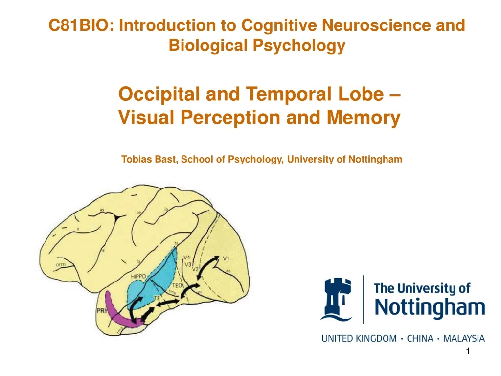
C81BIO: Introduction to Cognitive Neuroscience and Biological Psychology Occipital and Temporal Lobe – Visual Perception and Memory Tobias Bast, School of Psychology, University of Nottingham
The primary visual pathway includes: • Retina, lateral geniculate body, primary visual cortex. • Retina, lateral geniculate body, primary visual cortex, hippocampus. • Retina, superior colliculus, primary visual cortex.
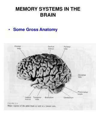
Outline • How does the brain process visual information beyond V1, and how does such information processing give rise to perception and memory? • Focus on: occipital lobe and temporal lobe (inferior temporal lobe and medial temporal lobe)
Hierarchy and functional differentiation in visual information processing Processing of visual information by the brain is hierarchical, with the complexity of the visual representation increasing from retina to visual association cortices and beyond. At the different stages of information processing there is functional differentiation, with different neuron types or different brain regions processing different properties of visual stimuli. Complex visual representations for perception and memory: • Integrated information concerning form, surface (colour, texture), spatial relationships, and movement • Integration with other sensory modalities (multimodal representations) Combination and elaboration via parallel channels Simple features: -Light intensity and wavelength -2D position in visual field
Visual information processing in extrastriate cortex V5/MT Posterior Parietal ctx. Superior colliculus V3 V3A STS eye LGN V1 V2 V4 TEO (posterior IT) TE (anterior IT) Extrastriate/prestriate ctx Inferior temporal (IT) ctx Occipital lobe
Visual processing in extrastriate cortex Neurons in extrastriate cortex signal ‘global’ properties of visual scenes and objects, rather than ‘component’ properties. Zeki S (2005) The Ferrier Lecture 1995. Behind the seen: The functional specialization of the brain in space and time. Phil. Trans. Roy. Soc. B 360:1145-1183.
Global colour vs. component wavelength • Perceived colour of an object depends not only on the wavelength reflected by object, but also on wavelength reflected by the surroundings (colour constancy; e.g., perceived colour of object does not change when viewed during sunset). • Some neurons in V4 are ‘colour’-sensitive (i.e., respond to wavelengths in the centre of their receptive field, depending on the wavelengths reflected from the background), whereas neurons in primary visual pathway and V2 are only ‘wavelength’-sensitive. http://www.thenakedscientists.com/HTML/articles/article/martinwestwellcolumn9.htm/
Global/pattern motion vs. component motion Taken from: Zeki S (1993) A vision of the brain. Blackwell Science Publications.
Two visual information processing streams Following V1 (and perhaps earlier) visual information processing seems to be mediated by two streams, that are anatomically and functionally differentiated. Dorsal stream Dorsal stream: Visuo-spatial (‘where’)/ visuo-motor (‘how’) processing V5/MT Posterior Parietal ctx. Ventral stream Superior colliculus V3 V3A STS Ventral stream: Object analysis (‘what’) eye LGN V1 V2 V4 TEO (posterior IT) TE (anterior IT) Extrastriate/prestriate ctx Inferior temporal (IT) ctx Occipital lobe
Visual Streams – what/where Inferior temporal lobe lesions (‘ventral stream’) in macaques impair object- discrimination/recognition (‘what’), but not object location (‘where’). Posterior parietal lesions (‘dorsal stream’) impair object location (‘where’), but not discrimination (‘what’). Mishkin M, Ungerleider LG, Macko KA (1982) Object vision and spatial vision: two cortical pathways. Trends Neurosci. 6:414-417.
Visual Streams – what/how Milner and Goodale proposed that the ventral stream processes visual information for object perception (‘what’), whereas the dorsal stream processes visual information for visuo-spatially guided action (‘how’). Key evidence: patients with occipito-temporal brain damage show severe forms of visual agnosia (i.e., deficits in aspects of visual perception without blindness), but intact visually guided actions, whereas patients with posterior-parietal lobe lesions show optic ataxia (i.e., deficits in visually guided reaching) with otherwise relatively intact visual function. For example, patient DF with extensive bilateral ventral-stream lesions has profound visual agnosia, but shows intact visually guided reaching: DF can act on visual stimulus (e.g., visuomotor posting), but is unable to make perceptual judgements (e.g., perceptual orientation matching) Milner AD, Goodale MA (1998) The visual brain in action. Psyche 4(12) http://psyche.cs.monash.edu.au/v4/psyche-4-12-milner.html
What does the observation of optic ataxia in a patient with posterior parietal lobe lesions suggest? • The posterior parietal lobe is required for object perception. • The posterior parietal lobe is required for visuo-spatially guided action. • None of the above.
Two visual information processing streams Dorsal stream Dorsal stream: Visuo-spatial (‘where’)/ visuo-motor (‘how’) processing V5/MT Posterior Parietal ctx. Ventral stream Superior colliculus V3 V3A STS Ventral stream: Object analysis (‘what’) eye LGN V1 V2 V4 TEO (posterior IT) TE (anterior IT) Extrastriate/prestriate ctx Inferior temporal (IT) ctx Occipital lobe
Visual perception and memory in inferior temporal cortex Neuron in TE responds to fractal shape in i regardless of size, orientation, and colour • The inferior temporal cortex receives inputs from extrastriate cortex and forms the final stage in the visual processing hierarchy of the ventral stream. • Neurons in the inferior temporal cortex can respond very selectively to specific shapes and objects. • These responses can show: -invariance to changes in size, orientation, and other properties – i.e., the neuron ‘recognizes’ object regardless of the viewpoint. -sustained activity in absence of visual object, reflecting short-term object memory Sustained response during retention delay on matching-to-sample task Other fractal shapes fail to trigger strong response Retention delay (16 s) Miyashita Y, Chang HS (1988) Neuronal correlate of pictorial short-term memory in the primate temporal cortex. Nature 331:68-70.
Face cells • Some neurons in the inferior temporal lobe show highly selective responses to individual faces. • The highly selective properties have been compared to those of ‘gnostic units’ or ‘grandmother neurons’, i.e. hypothetical neurons at the end of a processing hierarchy that ‘recognize’ individual entities, such as your grandmother (although face cells typically respond to several faces; also compare Quian Quiroga, 2016, Neuropsychologia, concerning an evaluation of the ‘grandmother’ neuron concept). • Areas showing selective responses to faces have also been identified in the human inferior temporal lobe using functional imaging (e.g., Fusiform Face Area) (Kanwischer N, Yovel G, 2006, Phil. Trans. R. Soc. B 361:2109). PS DP PS DP Perret DI, Mistlin AJ, Chitty AJ (1988) Visual neurones responsive to faces. Trends Neurosci. 10:358-364.
The Medial Temporal Lobe (MTL): Further processing of visual information and multimodal integration MTL Hippocampus • Ventral Dorsal Streams • Other sensory information (auditory, olfactory, gustatory, somatosensory, etc. • MTL is at end of visual-processing hierarchy, combining inputs from ventral and dorsal stream, and receives additional inputs from other sensory modalities. • It is thus in position to elaborate visual representations further and to generate multi-modal representations. • Examples of complex representations mediated by MTL structures include: • Complex spatial representations, requiring the encoding of relations between many visual stimuli. • Multimodal representations of experiences (‘episodic’ memory) and facts (‘semantic’ memory) (together referred to as ‘declarative’ memory).
What does neuroanatomy indicate about the MTL: • MTL receives only visual information, but highly processed. • MTL receives visual, auditory, olfactory, and other sensory information. • MTL should only respond to visual stimuli. • Both a) and c) are correct.
Patient H.M. Surgical removal of hippocampus and of parts of the surrounding cortices to stop epileptic seizures. Henry G. Molaison 1926-2008 • Following surgery, HM showed severe and pervasive deficit in remembering new and recent experiences, facts, and places, whereas other cognitive functions, including procedural learning, were largely intact. • These findings triggered enormous research activity on function of hippocampus and surrounding cortices. Corkin (2002) Nature Neurosci Rev 3:153
Selective place learning deficits after hippocampal lesions in rats Hippoc. lesion Cortical lesion Control Watermaze Representative swim paths on trial 28 Trials Hippocampal lesion Search preference for target region during 'probe’ trials ( ) Target region RGM Morris et al (1982) Nature 297:681
Hippocampal place cells ‘Place cells’ in rat hippocampus ‘Place cells’ in human hippocampus during virtual navigation J O´Keefe (2014) Nobel Lecture: Spatial Cells in the Hippocampal Formation www.nobelprize.org/nobel_prizes/medicine/laureates/2014/okeefe-lecture.html Nobel Prize in Physiology and Medicine 2014 AD Ekstrom et al (2003) Nature 425:124
Encoding of multimodal percepts by hippocampal neurons Hippocampal neuron with multimodal responses to Oprah Winfrey Quian Quiroga et al. (2009) Explicit encoding of multimodal percepts by single neurons in human brain. Curr. Biol. 19:1308-11313. (Also compare: Quian Quiroga R (2016) Neuronal codes for visual perception and memory. Neuropsychologia 83:227-241
Conclusion • Perception and memory based on visual (and other sensory) information can be understood as a hierarchically organized sequence of processing steps mediated by interconnected brain networks. • At the earliest stages neurons respond to very basic features. • At progressively higher stages, neurons respond to combinations of basic features and get activated by more and more complex stimuli. • Visual information processing is also characterized by functional differentiation, i.e. different properties of visual stimuli are processed in parallel by different neuron types/brain regions (e.g., colour and motion; information concerning stimulus identity vs. information relevant to what to do with a stimulus).
Occipital and temporal lobe: visual perception and memory – Selected Reading Textbook chapters: Carlson NR (any recent edition) The physiology of behavior. • Vision (Chpt. 6) • Relational learning and amnesia (Chpt. 15) Review articles: Mishkin M, Ungerleider LG, Macko KA (1982) Object vision and spatial vision: two cortical pathways. Trends Neurosci. 6:414-417. Quian Quiroga R (2016) Neuronal codes for visual perception and memory. Neuropsychologia 83:227-241.
Occipital and temporal lobe: visual perception and memory – Revision questions • What could be considered overarching principles of visual information processing? • Can you illustrate these principles based on examples from visual information processing along the primary visual pathway and beyond? • What are the ventral and dorsal visual streams? Which empirical evidence supports the existences of these two visual processing streams? • Describe the firing characteristics of neurons in the inferior temporal lobe that are selective for specific visual shapes and objects, or for faces. • What happens with visual information in the medial temporal lobe? • Describe some types of neurons in the hippocampus, with highly selective firing properties.
Occipital and temporal lobe: visual perception and memory – some further questions to ponder • What is a ‘colour’? • Can you explain why the right bottom corner of the two pictures is perceived as red in the left image, even though it reflects light of the same wavelength composition as the right bottom corner of the right image? • Can you explain, in principle, how we may recognise objects, faces, and places? • Can you think of differences between receptive fields of visual neurons and place fields of hippocampal neurons? • How could the brain mediate the ‘use’ of perception and memory to guide motor actions or their influence on emotions?