Download
1 / 33
370 likes | 651 Views
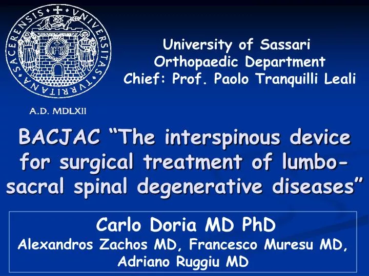
University of Sassari Orthopaedic Department Chief: Prof. Paolo Tranquilli Leali. BACJAC “The interspinous device for surgical treatment of lumbo-sacral spinal degenerative diseases”. Carlo Doria MD PhD Alexandros Zachos MD, Francesco Muresu MD, Adriano Ruggiu MD.

E N D
University of Sassari Orthopaedic Department Chief: Prof. Paolo Tranquilli Leali BACJAC “The interspinous device for surgical treatment of lumbo-sacral spinal degenerative diseases” Carlo Doria MD PhD Alexandros Zachos MD, Francesco Muresu MD, Adriano Ruggiu MD
Lumbar spinal stenosis (LSS) and symptomatic Degenerative Disc Disease (DDD) are the most common indications for lumbar spinal surgery in the elderly population Laminectomy and Fusion
Degenerative Cascade • Disc degeneration • Bulging of the annulus • Facet joint hypertrophy • Thickening of the ligamentum flavum A higher incidence in peri and post menopausal women has been noted Contribute to narrowing of the spinal canal and/or lateral foraminal recesses radicular pain
Symptoms are typically exacerbated on extension of the spine, e.g on standing or walking when the canal is further narrowed, and alleviated on flexion Chung SS, Lee CS, Kim SH (2000) Effect of low back posture on the morphology of the spinal canal. Skeletal Radiol 29:217-223 Baastrup Disease Facet Joint Syndrome Baastrup Cl. : Le “lumbago” et les affections radiologiques des apophyses epìneuses des vertebres lombaires; de la I vertebre sacrèe et des parties interepineuses. J Radiol Electrol 1936; 20:78-93
Baastrup Disease Facet Joint Syndrome Chronic Low Back Pain
Which way ???? Failure of conservative treatment (drugs, physiotherapy and epidural injection) • Microsurgical decompression /Fusion • Disc Arthoplasty (DDD alone without LSS) • Arthrorisis • Interspinous spacers • Dynamic Pedicle Screw Fixation • PercuDyn System SURGERY
Currently, motion preservation and dynamic stabilization also are rapidly developing in spinal surgery. Devices for these treatments include a variety of new implant technologies developed to preserve, limit or enhance motion of the spine • Posterior dynamic stabilization systems • Nucleus replacement devices • Total Disc Replacement • Interspinous devices
INTERSPINOUS SPACERS The interspinous device does not replace microsurgical decompression in patients with massive stenosis and continuous claudication, but offer a save, effective and less invasive alternative in selected patients with moderate “soft” spinal stenosis and symptomatic DDD • Unload the facet joints • Restore foraminal height • Provide stability (extension) • Retension of posterior annulus • Stretch the ligamentum flavum • Disc regeneration ?????? Rationale Schnake KJ, Putzier M, Haas NP, Kandziora F (2006) Mechanical concepts for disc regeneration. European Spine Journal, 15 (suppl. 3), S354-S360
DIAGNOSIS for correct indications The most important inclusion criterium was a reproducible alleviation of symptoms (leg pain and lumbago) on flexion and exacerbation of symptoms in extension of the lumbar spine (Positional Claudication) Clinical • Dynamic X-rays (max flexion and extension) • CT scan 3-D reformatted for study of neuroforamina • Functional (Upright) MRI examinations were able to demonstrate the positional-dependent stenosis Radiological Pre-op extension Post-op extension Pre-op flexion Post-op flexion
Dynamic lumbar Stabilization with interspinous implants: the history 1986 The first interspinous device in Europe Titanium PEEK
PEEK ADVANTAGES * Biocompatibility * Biostability * Compatibility with diagnostic imaging * More elastic than titanium (reduce the risk of spinous process stress fractures)
BACJAC Interspinous Decompression S Y S T E M • Simple procedure • * self-deploying • Least invasive • * unilateral approach • * tissue sparing • * ligament preserving • Minimal Risk of Subsidence • * large contact area • * near-physiologic modulus
CLINICAL ADVANTAGES * Increases stiffness to the treated segment relieving low back pain due to degenerative diseases * Maintains physiological lordosis * Limits but does not eliminate movement in the treated segment (extension) * Limits the “domino effect” of degenerative disc disease so called “junctional disease” observed after fusion procedures * Possibility at L5-S1 level * Local anaesthesia
INDICATIONS Low-back pain that accompanies degenerative lesions of grades II, II and IV (Pfirmann Classification) in lumbar segments primarily in young, active adults in the following indications: * “Soft” central or lateral stenosis * Massive herniated disc * Recurrent herniated disc * Degenerative disc diseases at a segment adjacent to fusion * Symptomatic Modic I degenerative changes
CONTRAINDICATIONS * Grade V degenerative lesions in the MRI classification of Pfirmann * Osteoporosis * Spondylolisthesis * Spinous process insufficiency * Non-specific low back pain * Infection
Over-sized of implant with over-distraction and relative segmental kyphosis • Placement too posterior of implant with possible damage of supraspinous ligament and dislodgement PITFALLS
SURGICAL TECHNIQUE - 1 • Patient under general anaesthesia or local anaesthesia combined with mild sedation • Prone position on radiolucent table • A midline skin incision of 5 cm on the spinous processes of the manipulated level (C-arm fluoroscopy) • The supraspinous ligament is preserved • The paraspinal muscles were elevated from one side of the spinous processes (right side) to the level of the facets and laminae
SURGICAL TECHNIQUE - 2 • The small dilator is introduced parallel to the vertebral spine with the tip in the cranial direction on the right side of the spinous process and advanced through the interspinous ligament from right to left to create a 1-5 mm pilot hole or perforation • The perforation in the interspinous space should be as anterior as possible to minimise the risk of dislodgement of device • The dilator is removed and the tips of the sizing distractor are introduced to the pilot hole from the right side. A cycle of tension and relaxation should be repeated two or three times
SURGICAL TECHNIQUE - 3 • Once the spacer is mounted on the dedicated introducer it is positioned to the right side of the spinous process • Once the spacer is placed through the interspinous ligament is need to procede to its final housing by opening the wings into the left paravertebral side (self-deploying) anchoring itself
OUR EXPERIENCE 2008-2010 : 73 BacJac devices in 59 adult patients • Mean Age: 52 yrs (range 43-78) • Ratio male /female: 35/24 SINGLE LEVEL * L3-L4: 8 cases * L4-L5: 27 cases * L5-S1: 3 cases The size of implanted BACJAC was: * 8 mm: 3 cases * 10 mm: 43 cases * 12 mm: 21 cases * 14 mm: 5 cases * 16 mm: 1 case ° Small for L5-S1: 7 implants DOUBLE LEVELS * L3-L4 and L4-L5: 15 cases * L4-L5 and L5-S1: 3 cases THREE LEVELS * * L2-L3, L3-L4 and L4-L5: 2 cases * L3-L4, L4-L5 and L5-S1: 1 case * Not recommended by Pioneer
Pre-operative diagnosis • “SOFT” LUMBAR SPINAL STENOSIS • * central: 14 cases • * lateral: 21 cases • DISC HERNIATION : 11 cases • RECURRENT DISC HERNIATION: 4 cases • SYMPTOMATIC DEGENERATIVE DISC DISEASE: 15 cases • FACET JOINT SYNDROME (anaesthetic block): 5 cases • DISLODGEMENT OF DEVICE: 2 cases • POST-SURGICAL SYNOVIAL CYST: 1 case
Clinical Assessment Patients were clinically evaluated regularly before surgery and during a follow-up period of 18 months using the Visual Analogue Scale (VAS) (0-10 points) and the Oswestry Disability Index (ODI) Pre-operative average VAS: 7.3 (range 5.2-8.7) Pre-operative average ODI: 40.3 (range 32.6-48.7)
Radiographic analysis Dynamic and static radiographs were obtained before surgery and post-surgery at 1, 6 and 18 months Functional (Upright) MRI examen is for us a DREAM ……… Intervertebral disc height (a) (measured from endplate to endplate on AP Ferguson and LL X-rays (pre and post-operative) Interspinous distance(b) in standing position was also measured a b
Post-operative MRI was performed in some selected cases: MULTILEVEL DEVICES (3 LEVELS) COMPLICATIONS/ REOPERATIONS (2 CASES)
Clinical Results The VAS decreased at 3.1 (range 2.0-4.2) on post-operative period with highly significant (P < 0.005). This effect remained remarkably stable throughout the follow-up period of 18 months 7.3 4.2 2.5 2.0
Clinical Results Mean ODI score at 1 month post-operative was 21.5, mean ODI score at 12 months postoperative was 15.3 and mean ODI score at 18 months post-operative was 12.4 21.5 18.4 15.3 12.4
Clinical Results In our experience there were no significant differences in post-operative VAS and ODI score related to pre-operative diagnosis
Radiological Results The mean posterior disc height measured on AP and LL x-rays was 9.8 ± 2.1 mm pre-operatively and 10.4 ± 2.3 mm post-operatively Retension of posterior annulus and reopening of neuroforamina The post-operative Interspinous Distance increased a mean of 12% respectly to pre-operative data (0.9 cm pre-op versus 1.3 cm post-op) with unloading of facet joints
Discussion Low back pain is usually aggravated by extension and relieved by flexion Interspinous devices relieves intradiscal pressure and widen the neural foramen Adequate positioning of BACJAC require a deep insertion at the base of spinous process
BACJAC system have common mechanical actions such as distraction between adjacent spinous precesses and restriction of extension achieving posterior neural decompression Simple procedure with a short “learning curve” Minimally invasive technique: unilateral surgical approach and ligament preserving
Risk factors for failure • Female (poor muscle tone abdominal wall and paravertebral muscles) • Hyperlordosis with high obliquity of spinous process • Hypoplasia of spinous process (S1) • Insufficiency of supraspinous ligament * • Overweight “The so called suprasinous ligament in the lumbar spine is not a true ligament, but is a dissection artifact mainly formed by decussation across the midline of the fibres of the right and left lumbosacral fascia” * Heylings DJA (1978) Supraspinous and interspinous ligaments of the human lumbar spine. J Anat 125:127-131
Take home message BACJAC SYSTEM is a handly and minimally invasive device but outcomes are strictly related to correct indications and its use must be reserved in select patients with positional claudication in absence of classic contraindications as severe stenosis that require wide decompression