Download
1 / 110
1.16k likes | 1.51k Views
Motor Neurons, the Neuromuscular Junction, and Muscle. Cory Toth. University of Calgary Medical School Neurosciences Course. August 27, 2007. Objectives. Define muscle disease (myopathy) and discuss their clinical presentation Discuss common forms of muscle disease

E N D
Motor Neurons, the Neuromuscular Junction, and Muscle Cory Toth University of Calgary Medical School Neurosciences Course August 27, 2007
Objectives • Define muscle disease (myopathy) and discuss their clinical presentation • Discuss common forms of muscle disease • Review of the neuromuscular junction (NMJ) • Define myasthenia gravis and discuss their clinical presentation • Discuss other forms of NMJ disease
Objectives 6) Describe motor neuron diseases (MND) and discuss their clinical presentation 7) Describe how MND is diagnosed 8) What is available for the patient with MND?
The Motor Unit • The motor unit is a group of muscle fibers and the single motor nerve that activates the fibers
Peripheral Nerve Muscle Motor Neuron NMJ
Muscle Contraction 1) Peripheral nerve impulse is required, with the impulse transferred from an axon to the SARCOLEMMA of a muscle cell
Muscle Contraction 2) The impulse travels along the SARCOLEMMA and down the T-TUBULES. From the T-TUBULES, the impulse passes to the SARCOPLASMIC RETICULUM
Muscle Contraction 3) As the impulse travels along the Sarcoplasmic Reticulum (SR), the calcium gates in the membrane of the SR open. As a result, CALCIUM diffuses out of the SR and among the myofilaments.
Muscle Contraction • Calcium fills the binding sites in the TROPONIN molecules. As noted previously, this alters the shape and position of the TROPONIN which in turn causes movement of the attached TROPOMYOSIN molecule
Muscle Contraction 5) Movement of TROPOMYOSIN permits the MYOSIN HEAD to contact ACTIN 6) Contact with ACTIN causes the MYOSIN HEAD to swivel
Muscle Contraction 7) During the swivel, the MYOSIN HEAD is firmly attached to ACTIN. So, when the HEAD swivels it pulls the ACTIN (and, therefore, the entire thin myofilament) forward (Many MYOSIN HEADS are swivelling simultaneously with a collective effort)
Muscle Contraction 8) At the end of the swivel, ATP fits into the binding site on the cross-bridge & this breaks the bond between the cross-bridge (myosin) and actin. The MYOSIN HEAD then swivels back. As it swivels back, the ATP breaks down to ADP & P and the cross-bridge again binds to an actin molecule
Muscle Contraction 9) As a result, the HEAD is once again bound firmly to ACTIN. However, because the HEAD was not attached to actin when it swivelled back, the HEAD will bind to a different ACTIN molecule (i.e., one further back on the thin myofilament). This action continues…
in the muscle, away from the neuromuscular junction • the AP is again all-or-nothing How do we generate an action potential in skeletal muscle?
Recordings in the junction reveal local potential changes before a regenerative action potential is produced.
If we block the ability of the postsynaptic receptor channels to open, we can observe local currents, but no action potential. These local currents are called end plate potentials (epps).
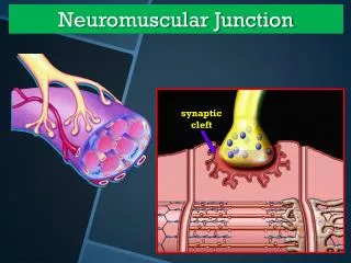
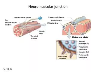
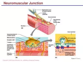
The Neuromuscular Junction (NMJ) The NMJ is an example of fast chemical transmission
The Neuromuscular Junction (NMJ) There are many ways that we manipulate the NMJ, or in which disorders manipulate the NMJ
Action potential Ca2+ channel Ca2+ Presynaptic terminal 1. An action potential arrives at the presynaptic terminal causing voltage gated Ca2+ channels to open, increasing the Ca2+ permeability of the presynaptic terminal.
- - + + + - - + - + - + - + + + - Look here + - + - + Neuromuscular Transmission: Step by Step Depolarization of terminal opens Ca channels Nerve action potential invades axon terminal
Ca2+ channel Presynaptic terminal Ca2+ ACh 2. Ca2+ enters the presynaptic terminal and initiates the release of a neurotransmitter, acetylcholine (ACh), from synaptic vesicles in the presynaptic terminal.
Synaptic cleft Na+ ACh Receptor molecule Na+ 3. Diffusion of ACh across the synaptic cleft and binding of ACh to Ach receptors on the postsynaptic muscle fiber membrane causes an increase in the permeability of ligand-gated Na+ channels.
ACh ACh Ca+2 ACh Ca+2 Na+ Na+ Na+ Na+ Na+ Na+ Na+ ACh Na+ Na+ Na+ Na+ Na+ Na+ Na+ Binding of ACh opens channel pore that is permeable to Na+ and K+. Ca+2 induces fusion of vesicles with nerve terminal membrane. ACh binds to its receptor on the postsynaptic membrane ACh is released and diffuses across synaptic cleft. Nerve terminal K+ K+ K+ K+ K+ Outside Muscle membrane Inside K+ K+ K+ K+ K+ K+ K+ K+
Na+ Action potential Action potential Na+ 4. The increase in Na+ permeability results in depolarization of the postsynaptic membrane; once threshold has been reached a postsynaptic action potential results.
EPP End Plate Potential (EPP) VNa The movement of Na+ and K+ depolarizes muscle membrane potential (EPP) 0 Muscle Membrane Voltage (mV) Threshold Presynaptic terminal -90 mV VK Time (msec) Presynaptic AP Outside Muscle membrane Inside ACh Receptor Channels Na Channels
ACh ACh receptor site Synaptic cleft Postsynaptic membrane Na+ 5. Once ACh is released into the synaptic cleft it binds to the receptors for ACh on the postsynaptic membrane and causes Na+ channels to open.
Acetic acid Choline ACh Acetylcholinesterase ACh receptor site 6. ACh is rapidly broken down in the synaptic cleft by acetylcholinesterase to acetic acid and choline.
ACh Acetic acid Synaptic vesicle Choline Choline ACh Presynaptic terminal 7. The choline is reabsorbed by the presynaptic terminal and combined with acetic acid to form more ACh, which enters synaptic vesicles.
Acetic acid Choline 8. Acetic acid is taken up by many cell types.
ACh Choline ACh ACh Choline ACh Acetate ACh Meanwhile ... ACh is hydrolyzed by AChE into Choline and acetate Choline resynthesized into ACh and repackaged into vesicle Choline is taken up into nerve terminal ACh unbinds from its receptor so the channel closes Nerve terminal Outside Muscle membrane Inside
The Neuromuscular Junction (NMJ) Structural Reality By John Heuser and Louise Evans University of California, San Francisco
The Neuromuscular Junction (NMJ) Structure-function of neurotransmitter postsynaptic receptors. 1. Nicotinic acetylcholine receptor of the neuromuscular junction. • composed of five subunits, composing a functional ligand-gated ion channel. • each subunit has four transmembrane spanning regions
The Neuromuscular Junction (NMJ) The molecules associated with the NMJ are numerous and complex – too much to know
The Neuromuscular Junction (NMJ) • Weakness occurs when the nerve impulse to initiate or sustain movement does not adequately reach muscle cells
ID: 29 yrs old RH Male CC: Abrupt onset of profound quadriparesis
Neuromuscular Presentation HPI: • Sore Muscles and felt fatigued after 40 minutes of working out in gym
Neuromuscular Presentation • Developed quadriparesis over next 20 hours • No sensory symptoms
Neuromuscular Presentation Review of Systems: • Denied numbness, pain, diplopia, dysarthria, dysphagia, bowel/bladder symptoms, shortness of breath. • Denies fever, rash, arthralgia, diarrhea, or vomiting prior to the onset.
Neuromuscular Presentation Past history • Denies past history of weakness • But had episode of feeling like “Jello” after working out in gym previously • Exercise-induced cramps, lasting over 2-3 days.