Download
1 / 55
550 likes | 601 Views
Explore the nature of white matter lesions in multiple sclerosis (MS), a chronic inflammatory demyelinating disease of the central nervous system. Learn about the pathology, symptoms, diagnosis, and clinical categories of MS.

E N D
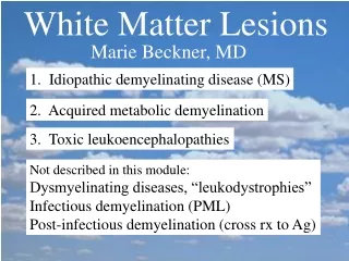
White Matter Lesions Marie Beckner, MD 1. Idiopathic demyelinating disease (MS) 2. Acquired metabolic demyelination 3. Toxic leukoencephalopathies Not described in this module: Dysmyelinating diseases, “leukodystrophies” Infectious demyelination (PML) Post-infectious demyelination (cross rx to Ag)
Primary Demyelination Damage to oligodendroglia & their myelin sheaths Axons are relatively preserved Secondary Demyelination Damaged axon loss of myelin Axonal transectionWallerian degeneration in distal portion
1. MULTIPLE SCLEROSIS First described in 1870’s Chronic, idiopathic, inflammatory demyelinating disease of CNS Selective destrx of oligodendrocytes & myelin with preserved axons Foci (plaques) widely dispersed in CNS ?Environmental influence acting upon genetically susceptible individuals
Multiple Sclerosis 1 million worldwide, increasing rate Higher prevalence in colder climates CNS lesions disseminated in space & time Symptoms: Paresthesias, gait difficulty, weakness/incoordination of 1 or both lower extremities, visual changes
MS - Magnetic Resonance Imaging MRI: T1, T2, FLAIR (fluid-attenuated inversion recovery) New lesions Gadolinium enhancement (recent disruption of blood brain barrier) Monitoring may help to identify agents that may be active against early inflammatory stage of MS
Hx: 25 yr woman with relapsing- remitting MS Axial FLAIR Periventricular hyperintense WM lesions NEJM 343:938-52, 2000
9 months later Axial FLAIR number & size of WM lesions NEJM 343:938-52, 2000
With gadolinium Many lesions demonstrate ring or peripheral enhancement NEJM 343:938-52, 2000
T1-weighted MRI Multiple regions of diminished signal, “black holes”, in peri- ventricular WM and corpus callosum. Chronic lesions of MS. NEJM 343:938-52, 2000
MS - Demyelinated Plaques Well-demarcated, gray, gelatinous S. Schochet
MS Plaque, often periventricular Lateral Ventricle Ellison & Love
MS Plaque Dawson’s fingers? Extensions along blood vessels Rarely see layers of demyelinated & more normally myelinated white matter (not here) Robbins, 6th ed.
Shadow Plaques - partial myelination adjacent to complete demyelination GRIPE
MS Plaque with H&E Stain Univ. Utah
MS Plaque Luxol Fast Blue Stain Univ. Utah
MS Plaque - Luxol Fast Blue Stain Univ. Utah
MS Plaque with Bodian Stain - Axons Univ. Utah
MS Demyelinated Plaques Loss of myelin (Luxol Fast Blue Stain) Perivascular lymphocytes Robbins, 6th ed., 2000
MS Demyelinated Plaques Preservation of axons Robbins, 6th ed., 2000
MS - Perivascular Lymphocytes Univ. Utah
MS-lymphocytes & reactive astrocytes Univ. Utah
MS-lymphocytes & reactive astrocytes Univ. Utah Enlarged, atypical nuclei, not hyperchromatic
MS Plaque - Subacute - Macrophages Univ. Utah
MS Plaque - Subacute - Macrophages Gitter cells - myelin breakdown products Univ. Utah
Creutzfeldt cell with minute chromatin fragments Often found in acute plaques of MS or in astrocytomas. Short-lived due to cell degen- eration. NEJM 339:542-9,1998
Multiple Sclerosis Other locations for plaques? Optic nerves, brain stem, cerebellum, spinal cord white matter, etc. What is Devic’s Disease? Demyelinating lesions of optic nerve(s) & spinal cord (neuromyelitis optica) Clinically, 30-40 yr, acute onset and often rapidly progressive
Devic’s Disease Optic Nerve Ellison & Love
Multiple Sclerosis - Tests IgG oligoclonal bands or IgG and lymphocytes (<50 cells) CSF: MRI: Abnormal in 95% patients Gadolinium enhanced lesions 5-10X > than clinical relapses Basis for future clinical trials as an outcome measure Abnormal evoked potential studies: central conduction velocities
MS Clinical Categories Relapsing-remitting - episodes of acute worsening w/ recovery & a stable course between relapses Secondary progressive - gradual neurologic deterioration w/ or w/o superimposed acute relapses in a patient who previously had relapsing-remitting MS Primary progressive- gradual, nearly continuous neurologic deterioration from the onset of sympt. Progressive relapsing- gradual neurologic deterioration from the onset of symptoms but with subsequent superimposed relapse
MS Variants - Clinical Progression Charcot type - most common (variable) relapsing-remitting - signs & symptoms w/n days recovery(wks) many develop secondary progression with persistent - signs of CNS dysfunction after relapse - disease may progress between relapses 10% benign MS - do well > 20 years 10% primary progressive MS - older patients, chronic progressive myelopathy Rare -Progressive relapsing MS
Rare MS Variants Acute MS (Marburg variant) Fulminant, rapid downhill course (fatal w/n months or 1 year) Younger patients Prominent tissue destruction in addition to demyelination CT & MRI lesions may be suspicious due to mass effect & edema and are then biopsied
Rare MS Variants cont... Schilder’s Disease (diffuse sclerosis) Bihemispheric widespread demyelination Scattered typical MS plaques Axonal injury, cavitation, & Wallerian degeneration with sparing of U fibers Leukodystrophy must be excluded Diff. Dx. Of “mental deterioration” in adolescents
Rare MS Variants cont... Balo’s Concentric Sclerosis Young patients Acute onset with strokelike symptoms Absence of remissions & exacerbations Affected tissue looks layered (onionskin) demyelinated white matter and more myelinated white matter (maybe due to remyelination)
2. Acquired Metabolic Demyelination Central pontine myelinolysis (CPM) Multifocal necrotizing leukoencephalopathy (MNL) Marchiafava-Bignami disease Osmotic demyelination syndromes ?
Central Pontine Myelinolysis What is the typical clinical scenario? Rapid serum Na+ in hyponatremic patient, (chronic liver dz, alcoholics) Very similar to MS plaque
Central Pontine Myelinolysis S. Schochet
Multifocal Necrotizing Leukoencephalopathy (MNL) Foci of necrosis with Ca++, WM > GM Formerly “focal pontine leukoencephalopathy” Predominantly immunosuppressed patients (AIDS, leukemia, irradiation, amphotericin B, methotrexate, other cytotoxic drugs) Clinically - complex neurologic abnormalities in patients with long critical illnesses
Multifocal Necrotizing Leukoencephalopathy Ellison and Love, Fig. 22.33, 1998 - ill-defined chalky white foci - distributed in pons and white matter
MNL - spongy vacuolation - swollen axons (may Ca++) - macrophages - foci often microscopic
Marchiafava - Bignami Disease Rare complication of alcoholism Destruction of myelinated fibers Degeneration of corpus callosum & anterior commissure Loss of callosal fiberscortical laminar sclerosis Morel’s laminar sclerosis (frontal & temporal cortex, usually layer III)
Marchiafava - Bignami Disease Ellison & Love
3. Toxic Leukoencephalopathy Variability: Diverse mechanisms Target of injury (myelin, astrocytes, blood vessels, etc.) Etiologies: Cranial irradiation Therapeutic drugs (antineoplastics) Drugs of abuse (cocaine, heroin, ecstasy or MDMA, toluene, etc.) Environmental solvents (organic solvents)
Targets of Toxins in Cerebral WM Filley & Kleinschmidt-DeMasters, NEJM, 2001
Toxic Leukoencephalopathies Clinical: WM tracts for higher cerebral function affected Inattention Forgetfulness Changes in personality Dementia Coma Death Absence of aphasia, language preserved
Cranial Irradiation More common for whole brain > local field Neurobehavioral dysfunction in 28% 3 stages: acute reaction - patchy, reversible WM edema delayed reaction - widespread edema & demyelination severe delayed reaction - loss of myelin and axons due to vascular necrosis and thrombosis
Chemotherapy Drugs Especially those given intrathecally Especially when given with irradiation Methotrexate - discrete or confluent necrosis - can cause MNL - vascular lesions in some Carmustine Others (cisplatin, cytarabrine, fluorouracil, levamisole, fludarabine, thiopeta) Sometimes IL-2, interferon-alpha
T2-MRI in man with right frontal GBM after radiation & chemotherapy with carmustine shows symmetric hyperintensity of the cerebral white matter (2 arrows) Filley & Kleinschmidt-DeMastsers, NEJM, p.428, 2001
Methotrexate Toxicity 6 yr old girl treated intrathecally for leukemia with CNS relapses