Download
1 / 26
2.04k likes | 7.1k Views
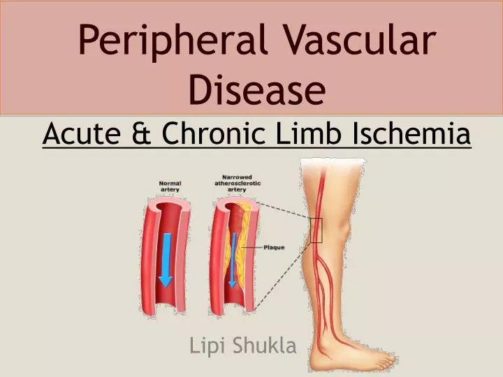
Peripheral Vascular Disease Acute & Chronic Limb Ischemia . Lipi Shukla . What is PVD?. Definition: Also known as PAD or PAOD. Occlusive disease of the arteries of the lower extremity. Most common cause: Atherothrombosis Others: arteritis, aneurysm + embolism.

E N D
Peripheral Vascular Disease Acute & Chronic Limb Ischemia Lipi Shukla
What is PVD? • Definition: • Also known as PAD or PAOD. • Occlusive disease of the arteries of the lower extremity. • Most common cause: • Atherothrombosis • Others: arteritis, aneurysm + embolism. • Has both ACUTE and CHRONIC Px • Pathophysiology: • Arterial narrowing Decreased blood flow = Pain • Pain results from an imbalance between supply and demand of blood flow that fails to satisfy ongoing metabolic requirements.
The Facts: • The prevalence: >55 years is 10%–25% • 70%–80% of affected individuals are asymptomatic • Pt’s with PVD alone have the same relative risk of death from cardiovascular causes as those CAD or CVD • PVD pt’s = 4X more likely to die within 10 years than pt’s without the disease. • The ankle–brachial pressure index (ABPI) is a simple, non-invasive bedside tool for diagnosing PAD — an ABPI <0.9 = diagnostic for PAD • Patients with PAD require medical management to prevent future coronary and cerebral vascular events. • Prognosis at 1 yr in patient’s with Critical Limb Ischemia (rest pain): • Alive with two limbs — 50% • Amputation — 25% • Cardiovascular mortality 25%
Risk Factors: • Typical Patient: • Smoker (2.5-3x) • Diabetic (3-4x) • Hypertension • Hx of Hypercholesterolemia/AF/IHD/CVA • Age ≥ 70 years. • Age 50 - 69 years with a history of smoking or diabetes. • Age 40 - 49 with diabetes and at least one other risk factor for atherosclerosis. • Leg symptoms suggestive of claudication with exertion or ischemic pain at rest. • Abnormal lower extremity pulse examination. • Known atherosclerosis at other sites (eg, coronary, carotid, or renal artery disease).
Chronic PVD History: • INTERMITTENT CLAUDICATION • Derived from the Latin word ‘to limp’ • “Reproducible pain on exercise which is relieved by rest” • Pain can also be reproduced by elevating the leg • “my legs get sore at night and feel better when I hang them over the edge of the bed” • Other Symptom/Signs: • A burning or aching pain in the feet (especially at night) • Cold skin/feet • Increased occurrence of infection • Non-healing Ulcers • Asymptomatic 3. Critical Stenosis = >60%, impending acute ischemic limb: - rest pain - ischemic ulceration - gangrene
30% Buttock & Hip Claudication ±Impotence – Leriche’s Syndrome Thigh Claudication 60% Upper 2/3 Calf Claudication Lower 1/3 Calf Claudication Foot Claudication
DDx of Leg Pain • Vascular • DVT (as for risk factors) • PVD (claudication) • Neurospinal • Disc Disease • Spinal Stenosis (Pseudoclaudication) • Neuropathic • Diabetes • Chronic EtOH abuse • Musculoskeletal • OA (variation with weather + time of day) • Chronic compartment syndrome
What does the ABI mean? CAUTION: Patient’s with Diabetes + Renal Failure: They have calcified arterial walls which can falsely elevate their ABI.
Investigations: • BLOOD TESTS: • FBE/EUC/Homocysteine Levels • Coagulation Studies • Fasting Lipids and Fasting Glucose • HBA1C • WHEN TO IMAGE: • To image = to intervene • Pt’s with disabling symptoms where revascularisation is considered • To accurately depict anatomy of stenosis and plan for PCI or Surgery • Sometimes in pt’s with discrepancy in hx and clinical findings • NON INVASIVE: • Duplex Ultrasound • normal is triphasic biphasic monophasic absent
ANGIOGRAPHY: • Non-invasive: • CT Angiogram • MR Angiogram • Invasive: • Digital Subtraction Angiography Gold Standard • Intervention at the same time
CT Angiography Digital Subtraction Angiography • Value of angiography • Localizes the obstruction • Visualize the arterial tree & distal run-off • Can diagnose an embolus: • Sharp cutoff, reversed meniscus or clot silhouette
Treatment: • 1. RISK FACTOR MODIFICATION: • Smoking Cessation • Rigorous BSL control • BP reduction • Lipid Lowering Therapy • 2. EXERCISE: • Claudication exercise rehabilitation program • 45-60mins 3x weekly for 12 weeks • 6 months later +6.5mins walking time (before pain) • 3. MEDICAL MANAGEMENT: • Antiplatelet therapy e.g. Aspirin/Clopidogrel • Phosphodiesterase Inhibitor e.g. Cilostazol • Foot Care
PCI/Surgery: • Indications/Considerations: • Poor response to exercise rehabilitation + pharmacologic therapy. • Significantly disabled by claudication, poor QOL • The patient is able to benefit from an improvement in claudication • The individual’s anticipated natural hx and prognosis • Morphology of the lesion (low risk + high probabilty of operation success) • PCI: • Angioplasty and Stenting • Should be offered first to patients with significant comorbidities who are not expected to live more than 1-2 years • Bypass Surgery: • Reverse the saphenous vein for femoro-popliteal bypass • Synthetic prosthesis for aorto-iliac or ilio-femoral bypass • Others = iliac endarterectomy & thrombolysis • Current Cochrane review = not enough evidence for Bypass>PCI • Amputation: Last Resort
Mr. X presents with an acutely painful leg: • You have had a busy day in the ED and the next patient to see is: • Mr. X – a 60 yr old gentleman with a very painful leg. • He tells you that he woke up this morning with an excruciating pain in his left leg and has never felt this pain before. MUST RULE OUT ACUTE LIMB ISCHEMIA ? Embolism (AF/Recent Infarct/Anuerysm) ? Thrombosis of native vessel or graft ?Trauma
Fixed mottling & cyanosis What are the features of an acute ischemic limb? • REMEMBER THE 6 P’S: • PAIN • PALLOR • PULSELESNESS • PERISHING COLD (POIKILOTHERMIA) • PARASTHESIAS • PARALYSIS
History & Exam Findings • Further Hx: • Smokes 20cigs/day for 30 years • 4 months of ‘leg cramps’ in BOTH legs • 2-3 weeks of intermittent chest palpitations • Has not seen a Dr. in the last month • Examination: • Inspection: • LLL: below the knee is pale/cool • Palpation: • Irregularly irregular pulse • LLL Capillary return is sluggish • No pulses palpable below L femoral artery • All pulses palpable but appear reduced in R leg • Normal Sensation + Movement bilaterally Impression? 60yo male with a L Acute Ischemic limb on the background of heavy smoking, untreated AF and symptomatic PVD.
What will you do now? • 1. CALL THE VASCULAR REGISTRAR • Simple measures to improve existing perfusion: • Keep the foot dependant • Avoid pressure over the heel • Avoid extremes of temperature (cold induces vasospasm) • Maximum tissue oxygenation (oxygen inhalation) • Correct hypotension • 2. ORDER INVESTIGATIONS • FBE • EUC • Coagulation Studies • Group and Hold • 12 Lead ECG • Chest XR • 3. INITATE ACUTE MANAGEMENT: • Analgesia • Commence IV heparin • Call Radiology for Angiography if limb still viable • Discuss with registrar: • Thrombotic cause ?cathetar induced thrombolysis • Embolic cause ?embolectomy • All other measures not possible Bypass/Amputation
Mr. X’s Complication • Angiogram is done in radiology • Shows acute thrombosis of L popliteal artery • Cathetar induced urokinase and heparin infusion is started • …. 3-4 hours later • Severe calf pain in the reperfused limb • All pulses are present • Leg is swollen, tense and +++ tender • REPERFUSION INJURY! • Restored blood flow can lead to unwanted local + systemic effects • 1) Washout = • Metabolic Acidosis • Hyperkalemia • ARF (myoglobinuria) • Non-cardiac APO • 2) Compartment Syndrome = • May need fasciotomy
Learning Outcomes Risk factors for PVD Recognise signs and symptoms of chronic ischemia of the lower limbs Differential diagnosis for leg pain Examine a chronic ischemic limb Understand medical/surgical of management of PVD Recognise an acute ischemic limb Know it is important to call the vascular registrar ASAP Know what investigations to order in the ED Be aware of the manifestations of reperfusion injury
References: Uptodate Articles: • Clinical features, diagnosis & natural history of lower extremity PAD • Treatment of chronic critical limb ischemia • Indications for surgery in the patient with lower extremity claudication • Norgren L, Hiatt WR, Dormandy JA, et al. Inter-Society Consensus for the Management of Peripheral Arterial Disease (TASC II). J Vasc Surg 2007; 45 Suppl S:S5 • McDaniel MD, Cronenwett JL. Basic data related to the natural history of intermittent claudication. Ann Vasc Surg 1989; 3:273. • Lane DA, Lip GYH. Treatment of hypertension in peripheral arterial disease. Cochrane Database of Systematic Reviews 2009, Issue 4. Art. No.: CD003075. DOI: 10.1002/14651858.CD003075.pub2 • Murabito JM, Evans JC, Nieto K, et al. Prevalence and clinical correlates of peripheral arterial disease in the Framingham Offspring Study. Am Heart J 2002; 143:961 • Peripheral arterial disease: prognostic significance and prevention of atherothrombotic complicationsPaul E Norman, John W Eikelboom and Graeme J HankeyMJA 2004; 181 (3): 150-154 • http://www.imagingpathways.health.wa.gov.au/includes/dipmenu/limb_is/summary.html