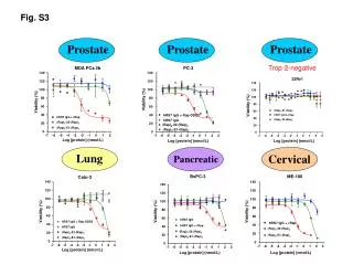
Cervical Incompetence
Cervical Incompetence. Dr.Sameera Madan. Definition. Inability of the uterine cx to retain a pregnancy in the absence of contractions or labor . Incidence and Prevalence. The lack of objective findings and clear dx criteria makes the incidence of CI difficult to ascertain . Risk Factors.

Cervical Incompetence
E N D
Presentation Transcript
Cervical Incompetence Dr.Sameera Madan
Definition Inability of the uterine cx to retain a pregnancy in the absence of contractions or labor Incidence and Prevalence The lack of objective findings and clear dx criteria makes the incidence of CI difficult to ascertain
Risk Factors Acquired Congenital • Trauma • Congenitally short Cx • Cx laceration following SVD • Mullerian duct abnormalities • Prolonged 2nd stage of labor • Deficiencies in Cx collagen & elastin • Surgical Procedures involving • the Cx (D&C, Cone bx) • Uterine over distention (multiple gestation, Polyhydramnios)
Clinical Manifestations ► The classic presentation of CI is: Cx dilatation and effacement in the 2nd trimester with fetal membranes visible at or beyond the external os in the absence of contractions. Which maybe asymptomatic or associated with: Vaginal fullness or pressure Spotting or bleeding An ↥ volume of watery, mucousy or brown VD Vague discomfort in the lower abdomen or back (sonographic manifestations of CI often occur prior to clinically detectable Cx changes)
Diagnosis Non-Pregnant women Pregnant women • There are no tests which can be performed to predict CI in a future pregnancy. • Those that have been proposed are inaccurate, inconvenient, or unproven in scientific studies • This may be based upon historical, clinical or sonographic criteria (non has been validated in well designed studies)
Pregnant women Historical features of CI • Hx of two or more 2nd trimester pregnancy losses • Hx of losing each pregnancy at an earlier gestational age • Hx of painless Cx diltation of up to 4-6 cm • Absence of clinical findings consistent with placental abruption • Hx of Cx trauma caused by • Cone bx • Intrapartum Cx lacerations • Excessive, forced Cx dilatation during TOP Harger JH. An evidance based analysis. Obstet Gynecol 2002;100:1313-27 (level II-2) Diagnosis
Pregnant women Clinical criteria • A digital examination should always be performed to evaluate the Cx in cases of CI, followed by TVS if the clinical examination is not diagnostic • Clinical findings include → significant premature Cx effacement and/or dilatation (> 2cm) specially with prolapse of fetal membranes into or completely through the endocervical canal (hourglassing) Diagnosis
Pregnant women Sonographic features • US of the Cx is more accurate and reproducible than D/E • The most consistent image of the Cx is obtained by TVS performed at ≥ 16 wks • Noninvasive stress techniques as : transfundal pressure, coughing and standing have been used to elicit US Cx changes • Serial US assessment of Cx length in women between 24-28 wks has been correlated with PTD Iams, JD, Goldenberg, RL, Meis, PJ, et al. N Engl J Med 1996; 334:567(level II-2) Diagnosis
Treatment approaches Non-surgical modalities Surgical (cerclage) • Modified activity • Bed rest • Pelvic rest • IM hydroxyprgesteron • Vaginal pessaries • Have not achieved wide spread acceptance and have not yet been proved to be effective The rational for surgery is that cerclage compensate for inherent Cx weakness & the only indication is the prevention of CI during pregnancy Cx cerclage is the standard treatment for CI, despite little data from randomized trials proving efficacy Newcomer J. Ostet Gynecol Surv 2000; 55:443-8. (level III) MRC/RCOG Working Party on Cervical Cerclage. Br J Obstet Gynaecol 1993; 100:516 (level I). Lazar, P, Gueguen, S, Dreyfus, J, et al. Br J Obstet Gynaecol 1984; 91:731. (level I)
Contraindications to Cerclage Maternal factors: • Premature labor • Premature rupture of membranes • Abruption of placenta • Intraamniotic, cervical, or vaginal infection • Medical condition that precludes administration of anesthesia or continuation of pregnancy Fetal factors: • Fetal demise • Fetal anomaly incompatible with extrauterine life • Nonreassuring fetal status • GA > 24 to 28 wks Treatment approaches Surgical
Cervical Cerclage Procedure Objective To reinforce the Cx at the level of the internal os; a 2ry effect is to lengthen the Cx Transvaginal Transabdominal Treatment approaches Surgical
Treatment approaches Surgical
Cervical Cerclage Transvaginal McDonald: a purse-string suture placed around the Cx as cephalad as possible & without dissection of the bladder or rectum. Treatment approaches Surgical
Cervical Cerclage Transvaginal Shirodkar: is performed using a 5 mm Mersilene tape placed around the Cx at the level of the internal os after surgically reflecting the UB anteriorly & the rectum posteriorly. Treatment approaches Surgical
Cerclage removal The cerclage is removed electively at 37 to 38 weeks or immediately with the onset of premature labor to avoid cervical laceration and/or uterine rupture Whether to remove the cerclage in the setting of PPROM is controversial A Shirodkar cerclage does not have to be removed if cesarean delivery is anticipated and future pregnancies are planned Treatment approaches Surgical