Download
1 / 1
20 likes | 164 Views
Results. Background. Aim. Subjects and Method. Conclusions. The effect of the alar base cinch suture in the treatment of Le Fort 1 osteotomy patients -a pilot study using a prospective randomised controlled trial study design Caitriona Howley, Shirley Cox.

E N D
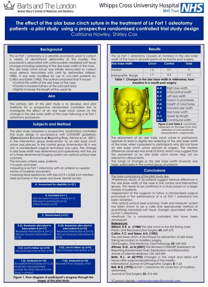
Results Background Aim Subjects and Method Conclusions The effect of the alar base cinch suture in the treatment of Le Fort 1 osteotomy patients -a pilot study using a prospective randomised controlled trial study design Caitriona Howley, Shirley Cox • The Le Fort 1 osteotomy is a versatile procedure used to correct a variety of dentofacial deformities of the maxilla. The procedure is associated with unfavourable nasolabial soft tissue changes including widening of the alar base width of the nose. • The alar base cinch suture was initially advocated to correct nasal defects associated with cleft lip deformities (Millard, 1980). It was later modified for use in non-cleft patients by Collins and Epker (1982). The authors proposed that it would:- • Control the width of the alar base of the nose • Create increased fullness of the subnasal area • Slightly increase the length of the upper lip The Le Fort 1 osteotomy caused an increase in the alar base width of the nose in all participants at six months post surgery. Table 1. Changes in the alar base width in millimetres, from baseline to 6 month post surgery • The placement of an alar base cinch suture in participants appears to lead to slightly less widening of the alar base width of the nose, when compared to participants who did not have an alar base cinch suture placed at surgery. The median difference observed was small (0.5mm) which may suggest that the placement of an alar base cinch suture may not be relevant in clinical terms. • The range of changes in the alar base width however was large, particularly in the control group of patients. • The main conclusions of this pilot study are: • Preliminary results of 20 patients suggest minimal difference in the alar base width of the nose in the control and intervention groups. This needs to be confirmed in a study based on a larger sample of patients. • Agreement of the surgeons to follow a standardised surgical procedure in the performance of a Le Fort 1 osteotomy has been obtained. • The optical surface laser scanning ‘mark and measure’ system has been shown to be a valid and reproducible method of quantifying nasolabial soft tissue changes associated with the Le Fort 1 osteotomy. • Methods for a randomised controlled trial have been developed. • References • Millard, D.R. Jr. (1980) The alar cinch in the flat flaring nose. • Plastic and Reconstructive Surgery 65: 669-672 • Collins, P.C. and Epker, B.N. (1982) • The alar base cinch: A technique for prevention of alar base flaring secondary to maxillary surgery. • Oral Surgery, Oral Medicine, Oral Pathology 53: 549-553 • Altman, D.G., et al (2001) The Revised CONSORT Statement for Reporting Randomised Trials: Explanation and Elaboration. • Annals of Internal Medicine 134: 663-694 • Betts, N.J., et al(1993) Changes in the nasal and labial soft tissues after surgical repositioning of the maxilla. • International Journal of Orthognathic Surgery 8: 7-23 • Bell, W.H. (1975) Le Fort 1 osteotomy for correction of maxillary deformities. • Journal of Oral Surgery 33: 412-426 • *Contact details: caitrionahowley@hotmail.com The primary aim of this pilot study is to develop and pilot methods for a prospective randomised controlled trial to investigate the effect of an alar base cinch suture on the change in the alar base width of the nose following a Le Fort 1 osteotomy procedure. Figure 2 and Table 2. Landmarks measured on the laser scan and definition of inter-landmark measurements respectively • The pilot study adopted a prospective randomised controlled trial study design in accordance with CONSORT guidelines; (Consolidated Standards of Reporting Trials, Altman et al., 2001). In the intervention group (intervention A) an alar base cinch suture was placed, in the control group (intervention B) it was not. A standardised surgical technique was used. The change in alar base width was determined post-operatively with the use of a three-dimensional imaging system (an optical surface laser scanner). • The inclusion criteria were, patients: • 16 years and older • requiring a Le Fort 1 osteotomy with an anterior or superior vector of maxillary movement • wearing fixed appliances with size 0.019 x 0.025 inch stainless steel archwires in the upper and lower dental arches A. Assessment for eligibility (n=22 ) B. Excluded (n=1 ) -Did not meet inclusion criteria (n=1) -Refused to participate (n=0) -Other Reasons (n=0) C. Randomised (n=21) D (i). Randomly allocated to intervention A (n=11) -Received intervention A (n=11) -Did not receive intervention A (n=0) D (ii). Randomly allocated to intervention B (n=10) -Received intervention B (n=10) -Did not received intervention B (n=0) E (ii). Lost to follow up (n=0) -Discontinued intervention (n=0) E (i). Lost to follow up (n=0) -Discontinued intervention (n=0) F (i). Analysed (n=10) Excluded from analyses (n=1) as treatment was performed outside the time interval for the study’s conduct F (ii). Analysed (n=10) Excluded from analyses (n=0) Figure 1. Flow diagram of participant’s progress through the stages of this pilot study