Periosteum
Periosteum. Periosteum. Central nervous system is protected by the bone structure (skull and vertebral column) as well as meninges. Meninges include dura mater, arachnoid and pia mater. These layers are continuous linings in both spinal cord and brain.

Periosteum
E N D
Presentation Transcript
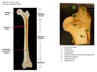
Periosteum • Central nervous system is protected by the bone structure (skull and vertebral column) as well as meninges. Meninges include dura mater, arachnoid and pia mater. These layers are continuous linings in both spinal cord and brain. • Periosteum, consists of collagenous connective tissue and arteries, covers the inner side of the skull. It also continues with the periosteum on the external surface of the cranium at the exit of foramen magnum and smaller foramina for nerves and blood vessels.
Periosteum • Periosteum and cranial bones are supplied by meningeal arteries. The largest is called middle meningeal artery, which splits into anterior and posterior branches after it enters the cranial cavity, supplies lateral surface of the cranium.
Dura Reflections • Dura Mater • Unlike dura mater in spinal cord, cranial dura mater is firmly attached to the periosteum. Subdural space, between the dura and arachnoid, is occupied by simple squamous epithelium and some fluid. • Dura reflections • The cranial dura is reflected along certain lines to form the dural reflections or dural septa
Dura Reflections • 1. The Falx Cerebri • a longitudinal fissure between the cerebral hemispheres. In front, it attaches to the crista galli of the ethmoid bone and goes back to tentorium cerebeli, hanging above corpus callosum • 2. The tentorium cerebelli • lies between occipital lobes and the cerebellum, the free border bounds incisura of the tentorium. • 3. The falx cerebelli • small dural fold in the posterior cranial fossa, extending vertically between the cerebellar hemispheres.
Transtentorial hernias • Expanding lesion of supratentorial compartment (tumor, hematoma) can push temporal lobe down into the incisura of the tentrium, causing impaired ipsilateral oculomotor nerve, first sign of this is impaired light reflex ipsilaterally. (dilated pupil). Further herniation can damage descending motor pathway causing upper neuron damage, with exaggerated reflexes (Barbinski’s Sign positive), either side or both (contralaterally)
Dural venous sinuses • Veins draining the brain empty into the venous sinuses of the dura mater, from which blood flows into the internal jugular veins. The wall of the sinus consists of dura mater, periosteum and endothelium • Dural venous sinus is formed by the outer periosteal and inner meningeal layer
Superior sagittal sinus • Lies along the attached border of falx cerebri • communication with nasal vein in the front • superior cerebral veins drain into it • continues with right transverse sinus
Inferior sagittal sinus • runs along the free border of falx cerebri • receives vein from medial aspects of the cerebral hemispheres • opens into straight sinus, which also receives great cerebral sinus • straight sinus usually continues with left transverse sinus • the sinus configuration around the internal occipital protuberance is referred as confluence of the sinuses.
Transverse sinus • Right (left) transverse sinus • lies in a groove on the occipital bone along the attached margin of tentorium cerebelli. • Becomes sigmoid sinus when reaches the petrous part of the temporal bone and continues with internal jugular vein
Other sinuses • The cavernous sinuses • on side of the sphenoid bone • receives the ophthalmic vein and the superficial middle cerebral vein • drains into transverse sinus via superior petrosal sinus
Other sinuses • inferior petrosal sinus • between the petrous part of the temporal bone and the basilar portion of the occipital bone • communication between the cavernous sinus and internal jugular vein • basilar sinus • connects cavernous and inferior petrosal sinus
Other sinuses • sphenoparietal sinus • small venous channel under the lesser wing of the sphenoid bone • drains into cavernous sinus
Emissary veins • connect dural sinuses with veins outside the cranial cavity • blood may flow either way
Pia and arachnoid layer • leptomeninges (slender membranes) : pia mater and arachnoid • The arachnoid contains fibroblasts, collagen fibers, and some elastic fibers
Subarachnoid cisterns • regions of subarachnoid space that contain substantial amounts of cerebrospinal fluid (CSF)
CSF • Production • generated mainly by the choroid plexuses of the lateral (largest and most important), third and fourth ventricles. • Choroid plexuses are formed by vascular pia mater
Function of CSF • Shock Absorbtion. Because the brain and spinal cord are suspended within the CSF, it cushions the CNS and protects it from traumatic injury. • Nutrition. The CSF contains sugars and other elements that are used by central nervous system cells, specifically neurons and glial cells. • Waste disposal. The CSF removes waste products produced by the metabolism of the cells in the CNS. • Communication. The CSF also acts as a messaging medium. Because the CSF contains a lot of active biochemicals (cytokines , hormones, neurotransmitters, metabolites and the like) the CSF provides information about the state of the CNS, whether it is running normally or whether there are any infections or dysfunctions.
CSF Circulation • lateral ventricles => through interventricular foramina to third ventricle => via cerebral aqueduct to fourth ventricle
CSF Circulation • From forth ventricle median aperture into cerebellomedullary cistern (cistern of magna); lateral aperture into pontine cistern interpeduncular cistern cistern of optic chiasma cistern of lamina terminalis cistern of corpus callosum
CSF absorption • Main site • Arachnoid villi that project into the dural sinus
CSF properties • Volume : 80 - 150 ml • Pressure: 80 - 180 cmH2O (recumbent, higher in lumbar area when sitting) • Clear and colorless, few cells (lymphocytes > 10 disease) • Glucose: half of plasma • Protein: very low
Hydrocephalus • Excess CSF • External: CSF in subarachnoid space • Internal: enlarged ventricles