Download
1 / 14
140 likes | 196 Views
This presentation outlines the challenges in retinal layer segmentation, proposes a graph-cut method for image segmentation, and discusses the results and conclusions. The motivation stems from combating leading causes of blindness like cataract, glaucoma, AMD, and diabetic retinopathy. Challenges in retinal diagnosis include unknown disease causes, manual segmentation errors, specialist variations, and inconsistent retinal structures. The proposed method involves image enhancement, gradient image computation, and building weights for segmentation, successfully segmenting 7 retinal layers across 8 boundaries. This automated approach improves segmentation accuracy and can aid in early disease diagnosis and medication monitoring.

E N D
Automated Layer Segmentation of Macula SD-OCT Images Using Graph-Cut Method Bashir I. Dodo, Yongmin Li, Khalid Eltayef and Xiaohui Liu Bashir Dodo (Bashir.dodo@brunel.ac.uk)
Presentation Outline • Motivation • Challenges in Retinal layer segmentation • Image Segmentation • Proposed Method • Result • Conclusion
Motivation The world’s four leading causes of blindness and visual impairment • Cataract -Affects the Lens and is noticed early by Patients • Glaucoma – Disease known for centuries, due to difficulties in its early diagnosis and frequent necessity of life-long treatment. • Age-related macular degeneration (AMD) - Ranks third among the global causes of visual impairment • Diabetic Retinopathy - Due to increase of Diabetics World Health Organization (WHO)
Challenges In Retinal Diagnosis Some challenges faced in the retinal layers segmentation for diagnosis are: • Causes of the diseases are unknown • Errors of manual segmentation • Variation from one specialist to another • Inconsistence of Retinal Structure • Speckle noise and shadows of blood vessel • Inexistence of universal Segmentation Algorithm
Image segmentation Image segmentation is the process of automating or facilitating the delineation of anatomical structures and other regions of interest(Pham, Xu, & Prince, 2000). Which is based on similarity, differences or proximity (Wertheimer, 1923). Figure 1: OCT image with boundaries and order of segmentation
Proposed Method: Schema Figure 2: Schematic representation of segmentation method
Proposed Method: Image Enhancement Top – Raw Images corrupt with noise Bottom – Enhanced images Figure 3: Unprocessed images (top) compared to pre-processed (bottom
Proposed Method: Gradient images Compute vertical Image Gradient Normalize Image Gradient Dark-light transition Get inverse of Image Gradient with *-1+1 Light-dark transition
Result Successfully segments 7 retinal layers across 8 boundaries. Applicable in real-time (~4.25sec per Image) Improved results accuracy Figure 4: Segmentation output- 7 layers across 8 boundaries
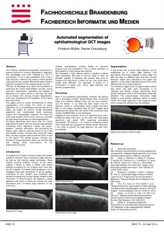
Result… The resolution of the B-scan images are 512 pixels in depth and 992 pixels across section with 16 bits per pixel. Ground truth images were labelled under the supervision of clinical experts.
Conclusion: Significance The importance of image segmentation in technology cannot be over emphasized as it plays a crucial role in many medical-imaging applications. Specifically, this study will aid in: • Preventing major eye diseases through early diagnosis • Monitoring progress of medication • Easing diagnostic process • Reducing variability in segmentation amongst professionals/ophthalmologists
CONCLUSION • Comparison with other methods • Reduce rigid nature of segmentation algorithm • Optimizing the flow of the gradient