Download
1 / 43
430 likes | 649 Views
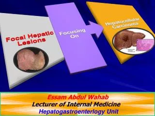
Essam Abdul Wahab Lecturer of Internal Medicine Hepatogastroenterlogy Unit. Objectives. Focal hepatic Lesions. Benign Liver Lesions. Hemangioma. The commonest liver tumor 5 % of autopsies Usually single small with demarcated capsule. Usually asymptomatic.

E N D
Essam Abdul Wahab Lecturer of Internal Medicine Hepatogastroenterlogy Unit
Hemangioma • The commonest liver tumor 5% of autopsies • Usually single small with demarcated capsule. • Usually asymptomatic. • US: well demarcated echogenic spot. • CT: venous enhancement from periphery to center • MRI: high intensity area • No need for FNA • No need for treatment ??
Benign nodule formation of normal liver with Central stellate scar. • Common in young and middle age women • No relation with sex hormones. • Asymptomatic, may cause minimal pain. • US: Nodule with varying echogenicity • CT: Hypervascular mass with central scar • MRI:iso or hypo intense • FNA: Normal hepatocytes and Kupffer cells with central core. • No treatment necessary. • Pregnancy and hormones----- OK.
Hepatic Adenoma • Benign neoplasm ---- normal hepatocytes but no portal tract, central veins, or bile ducts. • More common in women. • Usually asymptomatic but may have RUQ pain. • May presents with rupture, hemorrhage, or malignant transformation (very rare). • US: filling defect • CT: Diffuse arterial enhancement • MRI: hypo or hyper intense lesion • FNA : may be needed • Stop hormones, Observe every 6m If no regression then surgical excision.
Liver Cysts • May be single or multiple • May be part of polycystic kidney disease. • Patients often asymptomatic. • No specific management required. • Hydated cyst.
HBV carrier with HCC family history • Cirrhotic HBV carriers • Hepatitis C with cirrhosis • Stage 4 primary biliary cirrhosis • Genetic hemochromatosis and cirrhosis • Alpha-1 antitrypsin deficiency and cirrhosis • Other cirrhosis • 80% of patients with HCC have underlying cirrhosis Bruix J, et al. AASLD HCC guidelines. July 2010. Simonetti RS, et al. Dig Dis Sci. 1991;36:962-972.
Malignant Transformation HCC[2] Epigenetic alterations Genetic alterations Dysplastic nodules[1] Liver cirrhosis Hepatitis C,B Ethanol NASH Normal liver 1. Tornillo L, et al. Lab Invest. 2002;82:547-553. 2. Verslype C, et al. AASLD 2007. Abstract 24.
Contract enhanced US (CEUS) • Using microbubble structure, consisting of gas bubbles stabilized by a shell. • The sizes of the bubbles are not small to be lost by lung or large to pass to extracellular like CT and MRI.
Enhanced CT arterial phase Venous phase Delayed phase
Biopsy • Often not necessary. Most focal liver lesions have characteristic radiological findings on CT or MRI. • Recommended in uncertain cases. • Many debates regarding tunneling of tumor cells. .
HCC: Liver Transplantation • Best available treatment. • Removes tumor and liver. • Only if single tumor less than 5cm or less than 3 tumors less than 3 cm each. • Recurrence rate is low. • Not widely available.
HCC: Resection • Feasible for small tumors with preserved liver function (no jaundice or portal HTN) • Recurrence rate is high.
For non respectable pt. temporary measure only as bridge for liver transplantation. • For pt. with advanced liver cirrhosis: • Percutaneous Ethanol injection(PEI) • Radiofrequency ablation(RFA) • Tran arterial chemoembolization (TACE) • Microwave Coagulation.
Ethanol Injection It is the best known and best studied approach. It achieves necrosis rate of: • 90-100% of the HCC smaller than 2 cm • 70% in tumors between 2 and 3 cm • 50% HCC between 3 and 5 cm.
Radio Frequency Ablation • Deliver heat around the tip induces a wide region of tumor necrosis. • The efficacy of RFA in tumors <2 cm is similar to that of ethanol but requires fewer treatment sessions. The efficacy in tumors >2 cm is better than with ethanol .
Follow up • The efficacy of percutaneous ablation is assessed by dynamic CT or MR one month after therapy • The absence of contrast uptake within the tumor reflects tumor necrosis, while the persistence of contrast uptake indicates treatment failure