Thrombosis
Thrombosis. Dr. Nisreen Abu Shahin Assistant Professor of Pathology Pathology Department University of Jordan. Thrombosis. Pathogenesis (called Virchow's triad ): Endothelial* Injury ( Heart, Arteries) Stasis Blood Hypercoagulability

Thrombosis
E N D
Presentation Transcript
Thrombosis Dr. Nisreen Abu Shahin Assistant Professor of Pathology Pathology Department University of Jordan
Thrombosis • Pathogenesis (called Virchow's triad): • Endothelial* Injury ( Heart, Arteries) • Stasis • Blood Hypercoagulability * Endothelial cells are special type of cells that cover the inside surface of blood vessels and heart.
Contribution of Endothelial Cells to Coagulation • Intact endothelial cells maintain liquid blood flow by: 1- inhibiting platelet adherence 2- preventing coagulation factor activation 3- lysing blood clots that may form. • Endothelial cells can be stimulated by direct injury or by various cytokines that are produced during inflammation. • Endothelial injury results in: 1- expression of procoagulant proteins (tissue factor and vWF)local thrombus formation. 2- exposure of underlying vWF and basement membrane collagenplatelet aggregation and thrombus formation.
Response of Vascular Wall Cells to Injury(Pathologic effect of vascular healing) • Injury to the vessel wall results in a healing response, involving: -Intimal expansion (proliferating SMCs and newly synthesized ECM). this involves signals from ECs, platelets, and macrophages; and mediators derived from coagulation and complement cascades. - luminal stenosis & blockage of vascular flow
Causes of Endothelial injury • Valvulitis • MI • Atherosclerosis • Traumatic or inflammatory conditions • Increased Blood Pressure • Endotoxins • Hypercholesterolemia • Radiation • Smoking
Stasis • Stasis is a major factor in venous thrombi • Normal blood flow is laminar (platelets flow centrally in the vessel lumen, separated from the endothelium by a slower moving clear zone of plasma) • Stasis and turbulence cause the followings:
Causes of Stasis • Atherosclerosis • Aneurysms • Myocardial Infarction ( Non-cotractile fibers) • Mitral valve stenosis (atrial dilation) • Hyper viscosity syndromes (PCV and Sickle Cell anemia)
Hypercoagulability A. Genetic (primary): mutations in the factor V gene and the prothrombin gene are the most common B. Acquired (secondary): multifactorial and more complicated causes include: Immobilization, MI, AF, surgery, fracture, burns, Cancer, Prosthetic cardiac valves …etc
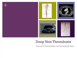
Morphology of thrombi • Can develop anywhere in the CVS (e.g., in cardiac chambers, valves, arteries, veins, or capillaries). • Arterial or cardiac thrombi begin at sites of endothelial injury; and are usually superimposed on an atherosclerotic plaque • Venous thrombi occur at sites of stasis. Most commonly the veins of the lower extremities (90%) • Thrombi are focally attached to the underlying vascular surface; arterial and venous thrombi both tend to propagate toward the heart. • The propagating portion of a thrombus is poorly attached fragmentation and embolus formation
ARTERY WITH AN OLD THROMBUS. A, H&e-stain. B, Stain For Elastic Tissue (black). The Original Lumen Is Delineated By The Internal Elastic Lamina (Arrows) And Is Totally Filled With Organized Thrombus.
lines of Zahn • Thrombi can have grossly (and microscopically) apparent laminations called lines of Zahn; these represent pale platelet and fibrin layers alternating with darker erythrocyte-rich layers. • Such lines are significant in that they represent thrombosis of flowing blood (can potentially distinguish antemortem thrombosis from postmortem clots) • postmortem blood clots are bland non-laminated clots (no lines of Zahn)
Mural thrombi = Thrombi occurring in heart chambers or in the aortic lumen. • Causes: -Abnormal myocardial contraction (e.g. arrhythmias, dilated cardiomyopathy, or MI) -endomyocardialinjury (e.g. myocarditis, catheter trauma) • Vegetations =Thrombi on heart valves 1- Bacterial or fungal blood-borne infections(infective endocarditis,). 2-Non-bacterial thrombotic endocarditisoccur on sterile valves.
Mural thrombi: A in heart; b in aortared arrows= mural thrombiblue arrows= lines of zhan
Fate of thrombi • Propagation Thrombi accumulate additional platelets and fibrin, eventually causing vessel obstruction • Embolization Thrombi dislodge or fragment and are transported elsewhere in the vasculature • Dissolution Thrombi are removed by fibrinolytic activity (Usually in recent thrombi) • Organization and recanalization -Thrombi induce inflammation and fibrosis. • recanalization(re-establishing some degree of flow) • Organization = ingrowthof endothelial cells, smooth cells and fibroblasts into the fibrin rich thrombus. 5. Superimposed infection (Mycotic aneurysm)
Venous thrombi • most common inveins of the legs • Superficial: e.g. Saphenous veins. - can cause local congestion, swelling, pain, and tenderness along the course of the involved vein, but they rarely embolize • Deep: e.g. Popliteal, Femoral and iliac vein. • more serious because they may embolize • can occur with stasis or hypercoagulablestates