Download
1 / 28
1.45k likes | 3.9k Views
Medical Parasitology Lecture notes. Actual live prep of Trichomonas vaginalis in DMEM with 10% serum under light microscope (400X). Dr. Paramita Basu. All About Protists. a polyphyletic collection of organisms most are unicellular

E N D
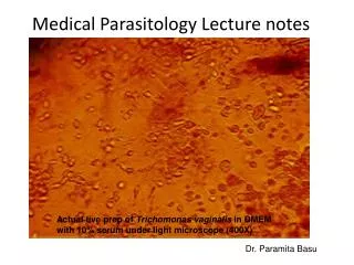
Medical Parasitology Lecture notes Actual live prep of Trichomonas vaginalis in DMEM with 10% serum under light microscope (400X) Dr. Paramita Basu
All About Protists • a polyphyletic collection of organisms • most are unicellular • lack the level of tissue organization present in higher eucaryotes • can be photoautotrophic, chemoorganotrophic, or mixotrophic • usually reproduce asexually by binary fission
Distribution of Protists • grow in a wide variety of moist habitats • most are free living • terrestrial and planktonic forms • parasitic forms cause disease in humans and domesticated animals
Types of Parasites • Protozoa(based on their life cycles and locomotion ) • Sarcomastigophora: Amebas (Sarcodina) & Flagellates (Mastigophora) • Ciliophora:Ciliates • Apicomplexans: (or sporozoa) • Helminths • Nemathelminthes: Roundworms • Platyhelminthes: Flatworms • Trematoda: Flukes • Cestoda: Tapeworms
Euglena. A Flagellate Pseudopodia. Ameba, Difflugia and Arcella have lobopodia; Chlamydophrys bears filopodia; Globigerina has reticulopodia.
Idealized (model) parasite life cycle and points of intervention. The human stages of the life cycle are located in the top half of the diagram. The extrahuman stages (in animate or inanimate reservoirs) are in the lower half. When the parasite reaches the infective stage, it invades the human host, matures, replicates, and ultimately completes the life cycle by producing infective forms. The infective forms are taken up by a vector or released into the environment. Control measures interfere with the replication or survival of the extrahuman stages of the parasite. They reduce the incidence of infection by reducing the number of infective stages to which humans are exposed. Immunization (vaccination) prevents symptomatic infection by inhibiting or killing the parasite as it enters (or replicates within) the human host. Chemoprophylaxis is used to inhibit parasite replication and thus prevent symptomatic infection. Neither immunization nor chemoprophylaxis prevents the initial entry of the parasite. Drug treatment is used to prevent death or severe morbidity in persons with established infections.
Parasite Survival Mechanisms in Immunologically Normal Hosts • Antigenic variation: trypanosomes, Plasmodium species, Giardia species • Intracellular infection: plasmodia, Toxoplasma species • Encystation: amebas, cestodes • Camouflage: schistosomes • Cleavage of antibodies or complement components: amebas, Leishmania species • Suppression or redirection of the cellular immune response: plasmodia, Leishmania species, schistosomes
Intestinal & Vaginal Protozoans • Intestinal protozoa are acquired by ingestion of cysts by the fecal–oral route of transmission, usually involving contaminated food or water. • Entamoebahistolytica lyses cells in the colon and feeds on their contents, resulting in colonic ulcerations and dysentery (diarrhea with blood and mucus). • Asymptomatic human carriers are the reservoir of infection; occasionally, the parasite enters the portal circulation and produces abscesses in the liver or other distant organs. • Giardialamblia and species of Cryptosporidium, Cyclospora, and Microsporidia are zoonotic and therefore cannot be prevented by control of human sanitation alone. They infect the small intestine and cause noninflammatory, watery diarrhea that can last for days or weeks. • A chronic inflammatory response to persistent giardiasis may result in the loss of intestinal villi, malabsorption syndrome, and weight loss. • Patients with AIDS and other immunodeficiencies may experience intractable diarrhea caused by these agents. • Trichomoniasis is a common form of sexually transmitted vaginitis and urethritis that requires treatment of all contacts to ensure that reinfection will not occur.
Intestinal Flagellates - Giardia lamblia Life cycle of Giardia lamblia. Humans acquire giardiasis by ingesting the cyst form of the parasite (1). After contact with the gastric contents, the parasite excysts and transforms to a trophozoite in the upper gastrointestinal tract (2), where it replicates asexually by binary fission (3). Trophozoites cause disease by attaching to the epithelium of the small intestine via a ventral sucking disk (4). As indicated by the solid line and open arrow below stage 6, trophozoites are not infectious for others because they are readily killed by drying in the external environment. As in amebiasis, humans acquire infection by ingesting cysts from the external environment (1), from the stools of other patients, or from their own stool (arrow from stages 5 to 2). Diagnostic Laboratory Tests - finding the distinctive cysts in formed stools, or cysts and trophozoites in liquid stools using ELISA Treatment - Metronidazole (Flagyl), Oral quinacrine hydrochloride (Atabrine) and furazolidone (Furoxone), Tinidazole (Fasigyn). Paromomycin (Humatin) may be useful in pregnancy
Vaginal Flagellate - Trichomonas vaginalis • Epidemiology - common inhabitant of the vagina and is found in 15% or more of women, in whom it occasionally causes vaginitis. Less common and less pathogenic species of are found in the gastrointestinal tract (T. hominis) or the mouth (T. tenax). • Spread -infection is transmitted by sexual intercourse. Infants may be infected during birth. • Diagnosis - Vaginal or urethral secretions or discharge should be examined microscopically in a drop of saline for characteristic motile trichomonads. • Treatment - Topical and systemic metronidazole (Flagyl) is best. • Tinidazole (Fasigyn) and ornidazole (Tiberal) are equally effective, • The patient's sexual partner should be examined and treated simultaneously. • Postmenopausal patients may require treatment with estrogens to improve the condition of the vaginal epithelium. • Prostatic infection can be cured with certainty only by systemic treatment with metronidazole or one of the above-mentioned nitroimidazoles.
Intestinal Amebas – Entameba histolytica Humans acquire amebic infection by oral ingestion of the cyst form of the parasite (1). Viable cysts may be ingested from the external environment (where they remain stable and infectious for prolonged periods after excretion), from the stool of other infected persons, or from the stools of the patients themselves (the arrow from 7 to 2). In the upper gastrointestinal tract, the parasite excysts after passing through the stomach (2), replicates asexually by binary fission (3), and transforms to the potentially pathogenic trophozoite form (4), which is typically found in the large intestine. Trophozoites die rapidly when they are shed into the external environment (5; solid line represents dead end). When conditions in the gastrointestinal tract are unfavorable, trophozoites transform into cysts (6–7), which can remain dormant for long periods of time in the host and the environment. • Pathology • cause destruction of host tissue (colon) • small ulcerations of the intestinal epithelium • Amebas within the lesions spread laterally in the deeper layers of the colon, sometimes producing flask-shaped ulcers in the mucosal epithelium. • Organisms may also spread through the portal circulation to produce abscesses in the liver, lung, brain, or other organs.
Intestinal Amebas – Entameba histolytica • Diagnostic Laboratory Tests • microscopic identification of trophozoites in freshly passed dysenteric stool or in scrapings from colonic ulcers obtained through a sigmoidoscope • stool immunoassay forantigen may be more sensitive than microscopic examination • serological diagnosis by IHA test • ultrasonography, CT, MRI, or radioisotope scanning • Treatment • Asymptomatic (cyst-passing) amebiasis can be treated with iodoquinol (Yodoxin), or diloxanide furoate (Furamide), or paromomycin (Humatin). • Metronidazole (Flagyl) is probably a drug of choice for symptomatic amebiasis • For mild to moderate intestinal disease, give metronidazole or tinidazole (Fasigyn) • For severe intestinal disease (amebic dysentery), give the regimen described above or, if the other regimens cannot be followed, dehydroemetine (or emetine).
Blood & Tissue protozoa • Most tissue invasive protozoa have intracellular stages in their life cycles. • Fever is a cardinal symptom of disseminated protozoal infections. Some species become dormant in tissues and cause prolonged, asymptomatic infection. • Malaria is transmitted among humans by Anopheles mosquitoes. Its propagation depends on the presence of a reservoir of partially immune, asymptomatic human carriers. Nonimmune persons (e.g., young children in endemic areas and adult travelers from nonendemic areas) may experience severe, or even fatal illness. • Because it is more virulent and more likely to be resistant to antimalarial drugs, Plasmodium faliciparum must be distinguished from other plasmodia and treated promptly. • Like malaria, babesiosis is a protozoal infection of erythrocytes, but it is more geographically localized and less prevalent, partly because it is transmitted by ticks rather than mosquitoes. • Virulence attributes of various Leishmania species determine whether infection results in a chronic skin ulcer at the site of the sand fly bite or a disseminated chronic febrile illness involving the liver, spleen, and lymph nodes. • Longstanding infection with the agent of Chagas disease, Trypanosoma cruzi, may result in immunopathologic damage to the heart and the gastrointestinal tract. • The agent of African sleeping sickness, Trypanosoma brucei, evades the host immune response by a genetically determined mechanism of antigenic variation.
Malaria • Genetic polymorphism of several human genes affects the entry, multiplication, and survival of malarial parasites and in determining the outcome of the infection. • Group A glycophorin and Duffy blood group antigen • Sickle cell Hemoglobin (Hbs) • Diagnostic Laboratory Tests • Microscopic examination of thick blood film stained with Giemsa's stain. • Examination of thin blood films stained with Giemsa's stain is necessary for species differentiation. • Several antigen-capture tests, using chromatographic methods to detect a trophozoite-derived protein in lysed blood, can be used for rapid diagnosis. They employ a dipstick or test strip with monoclonal antibodies against target parasite antigens. These rapid diagnostic tests (RDTs) can distinguish P falciparum or all four species but not the other three species individually. • Treatment & Prevention • Chloroquine (Aralen) is the drug of choice for treatment of malaria during the acute attack. • In cases of falciparum malaria coma (cerebral or algid malaria), parenteral quinine dihydrochloride or quinidine gluconate should be used until oral therapy is possible • Primaquine, an 8-aminoquinoline, eliminates the exoerythrocytic forms in the liver (potentially relapsing malaria), permitting a so-called radical cure. • Individuals deficient in glucose-6-phosphate dehydrogenase, frequently blacks or persons originally from the eastern Mediterranean, should be given a longer low-level course of primaquine (or none), owing to the possibility of hemolytic anemia • Drug-resistant strains of P falciparum should be treated with quinine sulfate plus a single dose of pyrimethamine-sulfadoxine (Fansidar), with quinine plus doxycycline or tetracycline, or with quinine plus clindamycin • Mefloquine (Lariam) and halofantrine (Halfan) are recommended alternative drugs for treatment of chloroquine-resistant P falciparum malaria.
Intestinal Helminths • The common intestinal helminthic infections are caused by nematodes (roundworms) and cestodes (tapeworms). • The life cycles of helminths require indiscriminate handling of human wastes leading to fecal helminth eggs contaminating soil, foodstuffs, animal feeds, and other materials. • Nematode infections are acquired by ingestion of eggs (e.g.,Ascaris, Trichuris, Enterobius ) or by direct penetration of soil larvae through the skin (e.g., , hookworm). These infections are often asymptomatic unless the worm burden is very large. • Pathologic features include intestinal obstruction (Ascaris), rectal prolapse (Trichuris), anal itching (Enterobius), and iron-deficiency anemia (hookworm). • Sustained autoinfection is a unique feature of strongyloidiasis. Immunosuppressed individuals may develop a syndrome of Strongyloids hyperinfection with diarrhea, pneumonitis, rash, and eosinophilia. • Diagnosis of intestinal helminths relies on identifying the characteristic eggs, larvae, or adult worms (or segments) in feces.
Tapeworms are long and ribbonlike and are composed of chains of rectangular segments (strobila), each segment bearing a complete male and female system, is capable of prodigious reproductive output. • There is no mouth and no trace of an alimentary system. Glucose or other simple predigested nutrients are absorbed directly from the host gut through millions of submicroscopic hair-like extensions, or microtriches, which interdigitate with the host's microvilli. • An highly muscular anterior holdfast organ (scolex) consisting of suckers and, anterior rings of muscle-controlled hooks maintains the worm's position in the gut or permits it to move freely in the small intestine. • All stages of tapeworms are parasitic. The adult is usually found in the intestine, whereas larvae develop in the tissues of various intermediate hosts, either vertebrate or invertebrate. • Life cycle
Blood & Tissue Helminths • Tissue-invasive helminths enter humans by ingestion (, or invasive cestodes), penetration through the skin (schistosomes), or arthropod bites (various filariae). • Humans usually acquire infection from undercooked meat from pigs or carnivorous game animals. Fever, severe muscle pain, and intense eosinophilia occur when larvae disseminate to human skeletal muscles. • Invasive cestode infections occur when humans ingest tapeworm eggs. Larvae that emerge from ingested eggs migrate to and encyst within the brain () or in the liver or lungs (). Symptoms develop after prolonged infection because of pressure effects of the enlarging cysts or hypersensitivity to released parasite antigens. • Schistosomiasis is prevalent in geographic areas where (1) fresh surface waters are used for bathing or washing, (2) waters are contaminated with human feces or urine, and (3) certain types of snails are present to host the intermediate stages of the parasite. • In humans, adult worms localize to the mesenteric or pelvic veins. The manifestations of disease are caused by the granulomatous reaction to helminth eggs that are trapped in tissues (e.g., intestine, bladder, liver). • Lymphatic filariasis results from chronic obstruction of lymph channels by adult worms and progressive swelling of extremities or genitals (e.g., elephantiasis). Adult worms are often inapparent in cutaneous filariasis, but their microfilaria (larvae) disseminate subcutaneously and produce chronic hypersensitivity reactions, which can lead to chronic dermatitis or blindness (e.g., onchocerciasis, or “river blindness”).
Comparative epidemiology of two tissue-invasive tapeworm infections: cysticercosisand echinococcosis (cystic hydatid disease) A. In pork tapeworm, the definitive host is the human, not the pig, because only humans harbor the adult tapeworms. Pigs are an intermediate host, since they develop only the tissue invasive form of infection (cysticercosis) after ingestion of eggs from feces of infected humans. Humans also develop the tissue-invasive stage of infection after ingesting eggs from human feces. Although tapeworm carriers can infect themselves with eggs from their own feces, many humans acquire cysticercosis directly by ingestion of fecally contaminated food and never actually harbor tapeworms themselves. B. The definitive host for the tapeworm that causes echinococcosis is the canine (dog, wolf, or fox). Intermediate hosts that harbor the tissue-invasive forms become infected by ingesting eggs from canine feces. In humans tissue-invasive disease with either tapeworm is considered a dead-end infection, since humans are rarely cannibalized (to complete the life cycle), and human remains are buried or incinerated in most cultures (making completion of the echinococcal life cycle improbable).
Helminths • Diagnosis • Ascariasis is readily diagnosed by stool examination using low-power (× 100) magnification. • Strongyloidiasis is diagnosed by identifying rhabditiform larvae in the stool usually accompanied by marked eosinophilia. • Tapeworm infections are readily diagnosed by stool examination. The proglottids are macroscopic and can be seen by the naked eye. • cysticercosis is usually diagnosed by its deep tissue manifestations (including lesions visible by CT scan or by a “soft” radiographic technique in the long axes of skeletal muscles). A positive serologic test for antibodies to is helpful, especially among persons who live in areas of low incidence. The test is typically negative in persons with intestinal infection only. • Most schistosome infections are diagnosed readily by microscopic examination of stool (S. mansoni, S. japonicum) or urine (S. hematobium) or by biopsy of a rectal valve (S. mansoni). • Infections that release microfilariae into the bloodstream (lymphatic filariasis) are diagnosed by examining smears of peripheral blood.
Helminths • Treatment • Mebendazole, albendazole, ivermectin, nitazoxanide, etc. are effective in treating Ascariasis • Albendazole, mebendazole, and pyrantel pamoate are used to treat hookworm infection • Albendazole and ivermectin are the drugs of choice for strongyloidiasis • Intestinal tapeworms are cured with a single dose of niclosamide • Praziquantel and albendazole are both effective in the treatment of cysticercosis. • Praziquantel is the treatment of choice for schistosomiasis. • treatment for lymphatic filariasis consist of diethylcarbamazine and ivermectin