Download
1 / 22
230 likes | 396 Views
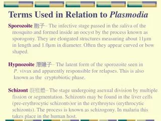
Figure 1.7 Regional terms used to designate specific body areas. 4. 1. 2. 5. 3. 6. 7. Figure 4.3a Epithelial tissues. 8. Figure 4.3b Epithelial tissues. 9. Figure 4.3c Epithelial tissues. 10. Figure 4.3d Epithelial tissues. 11. Figure 4.3e Epithelial tissues. 12.

E N D
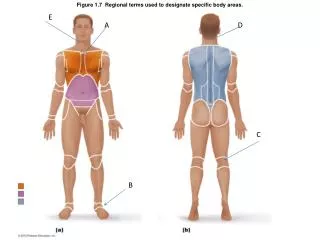
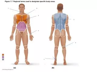
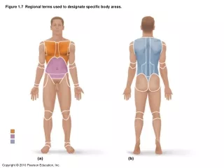
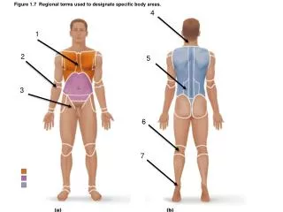
Figure 1.7 Regional terms used to designate specific body areas. 4 1 2 5 3 6 7
Figure 7.6a Inferior aspect of the skull, mandible removed. 16 21 17 18 22 19 20
Figure 7.31a The hip (coxal) bones. 23 26 24 27 25 28
Figure 10.5 Superficial muscles of the body: Anterior view. 32 29 33 30 31
Figure 10.12a Muscles of the abdominal wall. 34 37 35 36 38
Figure 12.10a Midsagittal section of the brain. 39 43 40 41 44 42 45
Figure 15.3a Extrinsic eye muscles. 47 46 48 49
Figure 15.24b Structure of the ear. 50 51 53 52
Figure 8.5d Movements allowed by synovial joints. 57 60 58 59
Sternal Antecubital Inguinal Nuchal Lumbar Popliteal Calcaneal Simple squamous epithelium Simple cuboidal epithelium Simple columnar epithelium Pseudo stratified epithelium Stratified squamous epithiliem Transitional epithelium Reticular connective tissue Hyoid bone Horizontal maxillary plate Zygomatic process Jugular foramen Magnum foramen External occipital protuberance Carotid canal Occipital condyles Iliac crest ASIS AIIS PSIS PIIS Acetabullum
Biceps brachii Gracilis Tibialus anterior Pectoralis major Brachioradialis Serratus anterior Transverse abdominis Internal oblique Rectus abdominis External oblique Intermediate mass of the thalamus Frontal lobe Pituitary gland Pons Arbor vitae Fourth ventricle Medulla oblongata Superior rectus muscle Lateral rectus muscle Medial rectus muscle Inferior oblique muscle Malleus Tympanic membrane Staples
Cochlea Circumduction Abduction Adduction Flexion Flexion Extension Extension