Download
1 / 44
440 likes | 465 Views
Explore the fascinating world of cells, the building blocks of life, and uncover their intricate structures and functions within the human body. Discover the essential components like plasma membrane, cytoplasm, and nucleus, as well as organelles such as mitochondria and lysosomes. Delve into the mechanisms of endocytosis, exocytosis, and protein synthesis. Embark on a journey to understand the dynamic nature of cells and their vital role in our existence.

E N D
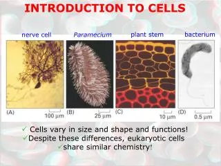
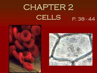
Introduction to Cells Cells – the smallest living units in our bodies There are approximately 70 TRILLION cells in our bodies! Each one is a separate life! Organelles – “little organs” – carry on essential functions of cells Enzymes – direct chemical reactions in cells Metabolism – the sum of all chemical reactions in the cell
Introduction to Cells Cells have three main components Plasma membrane Cytoplasm Nucleus
Structure of a Generalized Cell Figure 2.1
The Plasma Membrane Plasma membrane defines the extent of the cell Structure of membrane Fluid mosaic model (lipid bilayer) Types of membrane proteins Integral proteins – firmly imbedded in, or attached to lipid bilayer Peripheral proteins – attach to membrane surface
Our bodies are ONLY made of cells, extracellular fluids containing nutrients (a bunch of chemicals) and what is called extra cellular matrix which is manufactured by cells! Extracellular matrix includes, bone, tendons, ligaments, blood and other connective tissues. Cells are responsible for maintaining the composition of these tissues.
The Plasma Membrane Figure 2.2a
The Plasma Membrane Monday Functions – relate to location at the interface of cell’s exterior and interior Provides barrier against substances outside cell Some plasma membranes act as receptors
The Plasma Membrane • Determines which substances enter or leave the cell • Membrane is selectively permeable • Diffusion – molecules move from a region where they are more concentrated to an area where they are less concentrated • Osmosis – the diffusion of water across a membrane Membrane Structure
Endocytosis Endocytosis Mechanism by which particles enter cells Phagocytosis – “cell eating” Pinocytosis – “cell drinking” NOTE: Clathrin is a protein that causes bending of the cell membrane in order for a molecule to be captured by endocytosis Integral Proteins are involved in transport mechanisms across the plasma membrane
Receptor-mediated Endocytosis Receptor-mediated endocytosis Plasma proteins bind to certain molecules Invaginates and forms a coated pit Pinches off to become a coated vesicle NOTE: This is the method by which insulin and cholesterol enter cells!
Two Types of Endocytosis Figure 2.3
Exocytosis Exocytosis – a mechanism that moves substances out of the cell Substance is enclosed in a vesicle The vesicle migrates to the plasma membrane Proteins from the vesicles (v-SNAREs) bind with membrane proteins (t-SNAREs) The lipid layers from both membranes bind, and the vesicle releases its contents to the outside of the cell
Exocytosis This is how cells release hormones, enzymes and extra cellular matrix! Figure 2.4
The Cytoplasm Cytoplasm – lies internal to plasma membrane Consists of cytosol, organelles, and inclusions Cytosol (cytoplasmic matrix) Jelly-like fluid in which other cellular elements are suspended Consists of water, ions, and enzymes
Cytoplasmic Organelles Ribosomes – constructed of proteins and ribosomal RNA Site of protein synthesis NOTE: Most antibiotics work by blocking bacterial protein synthesis. The antibiotic works on one of the subunits to prevent bacteria from multiplying!
Cytoplasmic Organelles Endoplasmic reticulum – “network within the cytoplasm” Rough ER – ribosomes stud the external surfaces Smooth ER – consists of tubules in a branching network No ribosomes are attached; therefore no protein synthesis
The Endoplasmic Reticulum and Ribosomes Figure 2.5
Assembly of Proteins at the Rough Endoplasmic Reticulum Figure 2.6
Cytoplasmic Organelles Golgi apparatus – a stack of three to 10 disk-shaped envelopes Sorts products of rough ER and sends them to proper destination Products of rough ER move through the Golgi from the convex (cis) to the concave (trans) side
Role of the Golgi Apparatus in Packaging Products of Rough ER Figure 2.8
Cytoplasmic Organelles Lysosomes – membrane-walled sacs containing digestive enzymes Digest unwanted substances Peroxisomes – membrane-walled sacs of oxidase enzymes Enzymes neutralize free radicals and break down poisons Break down long chains of fatty acids Are numerous in the liver and kidneys
Mitochondria Mitochondria – generate most of the cell’s energy; most complex organelle More abundant in energy-requiring cells, like muscle cells and sperm Figure 2.10
Cytoplasmic Organelles Cytoskeleton – “cell skeleton” – an elaborate network of rods Contains three types of rods Microtubules – cylindrical structures made of proteins Microfilaments – filaments of contractile protein actin Intermediate filaments – protein fibers
Cytoskeleton: Microtubule Figure 2.11a
Cytoskeleton: Microfilament Figure 2.11b
Cytoskeleton: Intermediate Filament Figure 2.11c
Cytoplasmic Organelles Centrosomes and centrioles Centrosome – a spherical structure in the cytoplasm Composed of centrosome matrix and centrioles Centrioles – paired cylindrical bodies Consists of 27 short microtubules Act in forming cilia Necessary for karyokinesis (nuclear division)
Cytoplasmic Inclusions Temporary structures Not present in all cell types May consist of pigments, crystals of protein, and food stores Lipid droplets – found in liver cell and fat cells Glycosomes – store sugar in the form of glycogen
The Nucleus The nucleus – “central core” or “kernel” – control center of cell DNA directs the cell’s activities Nucleus is approximate 5µm in diameter
The Nucleus Figure 2.13
The Nucleus Nuclear envelope – two parallel membranes separated by fluid-filled space Chromatin – composed of DNA and histone proteins Condensed chromatin – contains tightly coiled strands of DNA
The Nucleus Chromatin – composed of DNA and histone proteins Extended chromatin – contains uncoiled strands of DNA DNA’s genetic code is copied onto mRNA (transcription) while in “extended chromatin” form Chromosomes – highest level of organization of chromatin Contains a long molecule of DNA
The Nucleus Nucleolus – “little nucleus” – in the center of the nucleus Contains parts of several chromosomes Site of ribosome subunit manufacture
Cellular Diversity Specialized functions of cells relates to Shape of cell Arrangement of organelles
Cellular Diversity Cells that connect body parts or cover organs Fibroblast – makes and secretes protein component of fibers Erythrocyte – concave shape provides surface area for uptake of the respiratory gases Epithelial cell – hexagonal shape allows maximum number of epithelial cells to pack together
Cells that Connect Body Parts or Cover Organs Figure 2.16, step 1
Cellular Diversity Cells that move organs and body parts Skeletal and smooth muscle cells Elongated and filled with actin and myosin Contract forcefully
Cells that Move Organs and Body Parts Figure 2.16, step 2
Cellular Diversity Cells that store nutrients Fat cell – shape is produced by large fat droplet in its cytoplasm Cells that fight disease Macrophage – moves through tissue to reach infection sites
Cells that Store Nutrients and Cells that Fight Disease Figure 2.16, steps 3–4
Cellular Diversity Cells that gather information Neuron – has long processes for receiving and transmitting messages Figure 2.16, step 5
Cellular Diversity Cells of reproduction Oocyte (female) – largest cell in the body Contains many copies of organelles for distribution to daughter cells Sperm (male) – possesses long tail for swimming to the egg for fertilization Figure 2.16, step 6
Cellular Death It is theorized that telomeres may program cell death. These molecules reside on the ends of chromosomes. Each time the cell divides, some of these are lost. At some point (after so many cellular divisions) a “stop division” signal is given and the cell dies. This is called cellular senescence.