Development of the Gut Tube and Heart in Mouse Embryos
220 likes | 323 Views
This overview presents the early embryonic development of the gut tube and heart in mice, focusing on the formation of body cavities and their respective mesodermal layers. It discusses how the endoderm folds to create the gut tube, while the somatopleure and splanchnopleure give rise to various organs and structures, including the heart. The progression from simple cardiac tubes to a functional heart with distinct chambers and vasculature, along with the significance of the yolk sac in hematopoiesis, is examined, highlighting developmental processes and congenital abnormalities.

Development of the Gut Tube and Heart in Mouse Embryos
E N D
Presentation Transcript
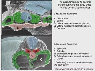
The endoderm folds to become the gut tube and the body sides form to enclose body cavities So S Sp A A N G E 9 day mouse, transverse A Split aorta G Gut tube So Somatopleure, parietal mesoderm* Sp Splanchnopleure, visceral mesoderm* C Cavity C So * Collectively a serous membrane around the body cavity Sp http://www.med.unc.edu/embryo_images/ 8 day mouse, transverse N Neural tube S Somite So Lateral mesoderm (somatopleure) Sp Lateral mesoderm (splanchnopleure) E Gut tube
The pleuropericardial cavity divides and the cavities are lined by parietal and visceral surfaces Aorta Oesophagus Lungs Peripleural cavity Heart Pericardial cavity Form a single serous membrane sac around the cavity Parietal surface of pleural and pericardial cavities Visceral surface of pleural and pericardial cavities
The peritoneal cavity is subdivided laterally at the rostral end but is a single cavity at the caudal end Aorta Pancreas Dorsal mesentery Stomach Small intestine Liver Ventral mesentery Form a single serous membrane sac around the cavity Parietal surface of peritoneal cavity Visceral surface of peritoneal cavity
DEVELOPMENT OF THE HEART AND GREAT BLOOD VESSELS LEARNING OUTCOMES 1. explain the early development of the heart from splanchnic mesoderm ahead of the neural plate which is then folded beneath the pharynx of the head fold. 2. outline the fusion of the endocardial tubes to form the simple linear heart with atrium, ventricle and valvular flaps pumping blood into the aortic arches. 3. define the three circulatory arcs of the heart supplying the body tissues, the yolk sac (vitelline) and the allantois and describe their functions 4. describe the role of the yolk sac splanchnopleure in early haematopoiesis 5. Understand the developmental process by which the aortic arches and truncus arteriosis are adapted to give the aortic and pulmonary trunks and the carotids 6. Show how septum formation in the primitive linear heart allows separate pumping of blood into the aorta and the pulmonary trunk 7. describe the congenital abnormalities of septal defects, patent ductus arteriosus, and persistent aortic arch
MESODERM INTERMEDIATE MESODERM TYPE STRUCTURE DERIVATIVE HEAD HEAD MUSCLES, SKULL, CARTILAGE CHORDA- NOTOCHORD LIMB MUSCLES AXIAL SKELETON PARAXIAL SOMITES TRUNK MUSCLES ICM EPIBLAST MESODERM DERMIS INTERMEDIATE PARTS OF KIDNEY AND REPRODUCTIVE TRACT LIMB SKELETON HEART LATERAL BODY CAVITY DIVIDERS BLOOD CELLS AMNION CHORION YOLK SAC ALLANTOIS FORMATION OF THE MAMMALIAN GASTRULA - 9
INNER CELL MASS EPIBLAST ENDODERM ECTODERM MESODERM Extraembryonic mesoderm of yolk sac and allantois Blood cells Splanchnic mesoderm Connective tissue, smooth muscle of viscera and blood vessels Heart Vascular endothelium * epithelial part only of organ Hair, nails Sweat glands Anal canal Extraembryonic endoderm of yolk sac and allantois Oral epithelium Lungs* Olfactory epithelium Mammaryglands Thyroid* Pharynx Surface epithelium Teeth enamel Gut tube* Stomodeal epithelium Pharyngeal pouches Liver* Anterior pituitary Allantois* Neural tube Pancreas* Head mesoderm Bladder* Middle Ear* Cranial motor nerves Paraxial mesoderm Lateral mesoderm Brain Skull Branchial cartilage Parathyroid* Spinal cord Notochord Intermediate mesoderm Sclerotome Spinal motor nerves Tonsils* Eye Somatic mesoderm Parietal pleura, pericardium, peritoneum Myotome Kidney and reproductive tracts Neural crest Axial skeleton Primary germ cells Dermatome Extraembryonic mesoderm of amnion and chorion Gonads* Cranial sensory nerves Visceral pleura Visceral peritoneum Mesenteries Dermis Trunk Muscles Teeth dentine Appendicular muscles Adrenal medulla Sympathetic ganglia Spinal sensory nerves Melanocytes THE MAP OF ORIGINS
B GUT TUBE CARDIAC TUBE VITELLINE VEINS The heart is a U-shaped tube at this stage and the forming blood vessels are initially unconnected The cardiac tube folds under the gut tube…… THE EARLY DEVELOPMENT OF THE HEART - 1 A DORSAL AORTA ENDODERM PERICARDIAL CAVITY CARDIAC PRIMORDIUM (SPLANCHNIC MESODERM) The cardiac primordia are established in the early gastrula as regions of splanchnic mesoderm ahead of the embryo itself. As a result of the head fold, this region ends up beneath the pharynx.
……and connects bilaterally with the dorsal aorta via the aortic arches C FUSED DORSAL AORTA ST 1 AORTIC ARCH (R) VENTRICLE ATRIUM VENOUS RETURN FROM CARDINAL VEINS, ORAL PLATE VITELLINE VEIN AND ALLANTOIC (UMBILICAL) VEIN The sides of the U-tube then fuse to produce the atrial and ventricle regions with valvular flaps to prevent back flow so that the heart can function as a simple peristaltic pump. The dorsal aorta form independently and then grow to meet the ventral output from the heart in the aortic arches
This pattern of mammalian development is a good example of recapitulation The diagram shows 6 aortic arches but, in mammals, 1 and 2 are regressing while the later arches are forming and arches 5 never form
REMINDER: The branchial arches and clefts and the juxtaposed pharyngeal pouches are a recapitulation of the respiratory anatomy of fish
Mouse, 8 day, sagittal Mouse, 9 day, frontal Mouse, 10 day, frontal The heart twists so that the atrium is rostral to the ventricle Mouse, 8 day, frontal The heart folds under the pharynx Mouse, 9 day, side http://www.med.unc.edu/embryo_images/
Cardinal veins Dorsal aorta Mesonephros Aortic arches Vitelline vein Vitelline artery Allantoic artery Allantoic vein Chorio-allantoic placenta Deoxygenated blood Mixed blood Yolk sac Oxygenated blood The embryonic circulation has three circulatory arcs through which blood is pumped by a simple linear heart
1. BODY CIRCULATION TRANSPORT OF O2 /FOOD MATERIALS TO TISSUES TRANSPORT OF WASTE MATERIALS AWAY 2. VITELLINE CIRCULATION CARRIES MOBILISED FOOD MATERIALS FROM THE YOLK SAC LOST FUNCTION IN MAMMALS BECAUSE SAC EMPTY CARRIES FIRST BLOOD CELLS FROM YOLK SAC SPLANCHNOPLEURE 3. ALLANTOIC CIRCULATION IN MAMMALS TAKES OVER THE FUNCTIONS OF THE VITELLINE ARC IN BIRDS SUPPLIES FOOD MATERIALS FROM MATERNAL CIRCULATION RETAINS AVIAN FUNCTION OF REMOVAL OF WASTE AND GAS EXCHANGE THE CIRCULATORY ARCS OF THE EMBRYONIC BLOOD SUPPLY
CELL CLUSTERS ENDOTHELIAL CELLS HAEMATOPOIETIC CELLS FORMATION OF BLOOD VESSEL AGGREGATION OF FURTHER MESENCHYME TO FORM MUSCULAR AND CONNECTIVE TISSUE WALL Haematopoiesis begins in the splanchnopleure of the yolk sac before transferring to the embryo itself later in development MESENCHYME IN SPLANCHNOPLEURE OF YOLK SAC
CAROTIDS(from L and RIII) AORTA (from LIV) I II III III RIGHT SUBCLAVIAN (from RIV) DUCTUS ARTERIOSUS (LVI to LIV) IV IV V VI VI PULMONARY TRUNK (from LVI) TA A RA LA A SEPTA V V LV RV VENOUS RETURN After birth venous return is from vena cava (blue arrows) and pulmonary veins (red arrows) The simple tubular heart twists to prepare for septum formation and the creation of a four-chambered organ. The aortic arches are selectively modifed to give rise to the great arteries THE HEART AND THE AORTIC ARCHES - FORMATION OF THE GREAT BLOOD VESSELS NOTES: 1. View from ventral surface 2. RA - Right atrium, LA - Left atrium, RV - right ventricle, LV - left ventricle, TA = truncus arteriosus
The separation between atria and between ventricles and between atria and ventricles occurs by means of septum formation
A Mouse, 12 days, section of truncus arteriosus Cushions form within the truncus arteriosus and will fuse to form the aortico-pulmonary septum separating the aortic and pulmonary flows Mouse, 10 days, frontal section Blood from the atrium passes to the ventricle by means of a channel. The beginnings of interatrial septum formation can be seen (A) http://www.med.unc.edu/embryo_images/
There is a split between deoxygenated blood returning from the rostral end of the foetus and oxygenated blood returning from the placenta. This spit is achieved by directed flow through the foramen ovale FOETAL CIRCULATION 25 Brachycephalic vessels 14 To 19 lungs DA From lungs FO 25 19 25 Liver 14 30 22 Trunk Placenta Hindlimb
CHANGES IN THE CIRCULATION AT BIRTH • Contraction of allantoic artery and veins to force placental blood • into main circulation. Rupture of umbilical cord • Contraction of Ductus arteriosus and closure of Foramen ovale so that right side blood is directed to lungs
INTER-VENTRICULAR SEPTAL DEFECT (Tetralogy of Fallot is variation on this) DEFECTIVE SEPTUM FORMATION INTER-ATRIAL SEPTAL DEFECT (persistent Foramen ovale)
R i g h t 4 L e f t 6 O e s o p h a g u s P E R S I S T E N T R I G H T A O R T I C A R C H I V P E R S I S T E N C E O F A O R T I C A R C H E S A N D V A S C U L A R R I N G A N O M A L I E S P A T E N T D U C T U S A R T E R I O S U S
REFERENCES Carlson BM (2003) Patten's Foundations of Embryology Noden DM, de Lahunta (1985) A Embryology of domestic animals McGeady TA, Quinn PJ, Fitzpatrick ES, Ryan MT (2006) Veterinary embryology University of North Carolina web site: http://www.med.unc.edu/embryo_images/