Download
1 / 62
630 likes | 923 Views
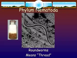
NEMATODA. NEMATODES. NEMA: thread EIDOS: form Nematohelminthes are either : round, cylindrical, spindle shaped. Examples: ENTEROBIASIS = OXYURIASIS ( pin worm) ASCARASIS = round worm ANCYLOSTOMIASIS = hook worm TRICHINOSIS TRICHURIASIS = whip worm.

E N D
NEMATODES NEMA: thread EIDOS: form Nematohelminthes are either : round, cylindrical, spindle shaped. Examples: • ENTEROBIASIS = OXYURIASIS ( pin worm) • ASCARASIS = round worm • ANCYLOSTOMIASIS = hook worm • TRICHINOSIS • TRICHURIASIS = whip worm
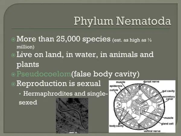
Ascaris lumbricoides • Giant intestinal roundworm • Whitish or pinkish worm • More than 1 billion individuals are affected • 70% from Asia • Soil transmitted helminth • Polymyarian • Somatic muscles are numerous and project well into the body cavity
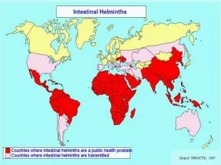
Epidemiology • World wide distribution, very common in China, especially in the countryside. Factors favoring the spread of the transmission: 1. Simple life cycle. 2. Enormous egg production ( 240,000 eggs/ day/ female ). 3. These eggs are highly resistant to ordinary disinfectants( due to the ascroside).The eggs may remain viable for several years. 4. Social customs and living habits. 5. Disposal of feces is unsuitable.
Ascaris lumbricoides • Males = 10-31 cm • Ventrally curved posterior end with 2 spicules • Females = 22–35cm • Straight, conical posterior end
Ascaris lumbricoides • Trilobate lips
The lips of Ascaris lumbricoides The three lips are seen at the anterior end. The margin of each lip is lined with minute teeth which are not visible at this magnification.
Ascaris lumbricoides • Unfertilized eggs • Narrower • Longer • Thin shell • Refractile granules are not organized • Found in the absence of males
Ascaris lumbricoides • Fertilized eggs • Thick transparent hyaline shell • Ovoid mass of protoplasm which develops into larvae at about 14 days
Ascaris lumbricoides • Embryonated eggs • Infective stage • Embryonation occurs in the soil
Life Cycle • 1.Site of inhabitation: small intestine 2. Infective stage: embryonated eggs 3. Route of infection: by mouth 4. No intermediate and reservoir hosts 5. Life span of the adult: about 1 year This worm lives in the lumen of small intestine, feeding on the intestinal contents, where the fertilized female lays eggs. An adult female can produce approximately 240,000 eggs per day, which are passed in faeces. When passed, the eggs are unsegmented and require outside development of about three weeks until a motile embryo is formed within the egg.
Ascaris lumbricoides Retrieved fromhttp://parasitol.wkhc.ac.kr/image/nema/lifeal.JPG Feb. 2008
Pathogenesis There are two phase in ascariasis: 1. The blood-lung migration phase of the larvae: During the migration through the lungs, the larvae may cause a pneumonia. The symptoms of the pneumonia are low fever, cough, blood-tinged sputum, asthma. Large numbers of worms may give rise to allergic symptoms. Eosionophilia is generally present. These clinical manifestation is also called Loeffler’s syndrome.
2. The intestinal phase of the adults. The presence of a few adult worms in the lumen of the small intestine usually produces no symptoms, but may give rise to vague abdominal pains or intermittent colic, especially in children. A heavy worm burden can result in malnutrition. More serious manifestations have been observed.
Complication • Wandering adults may block the appendical lumen or the common bile duct and even perforate the intestinal wall. Thus complications of ascariasis, such as intestinal obstruction, appendicitis, biliaryascariasis, perforation of the intestine, cholecystitis, pancreatitis and peritonitis, etc., may occur, in which biliaryascariasis is the most common complication.
Diagnosis • The symptoms and signs are for reference only. The confirmative diagnosis depends on the recovery and identification of the worm or its egg. A. Ascarispneumonitis: examination of sputum for Ascaris larvae is sometimes successful. B. Intestinal ascariasis: feces are examined for the ascaris eggs. (1) direct fecal film: it is simple and effective. The eggs are easily found using this way due to a large number of the female oviposition, approximately 240,000 eggs per worm per day. So this method is the first choice. (2) brine-floatation method: (3) recovery of adult worms: when adults or adolescents are found in feces or vomit and tissues and organs from the human infected with ascarids , the diagnosis may be defined.
Ascaris lumbricoides • Treatment • Albendazole • Drug of choice • 400 mg single dose • 200 mg for children under 2 year old • Mebendazole • 500 mg single dose • Pyrantel Pamoate • 10 mg/kg body weight (max. of 1 g)
Prevention • 1.Sanitary disposal of faeces. • 2.Hygienic habits such as cleaning of hands before meals. • 3.Health education.
Ancylostoma duodenale • Slightly larger than Necator americanus • Head curves in the same direction as the curvature of the body
Morphology 1. Adults: They look like an odd piece thread and are about 1cm. They are white or light pinkish when living. ♀is slightly larger than♂.The male’s posterior end is expanded to form a copulatory bursa. 2. Eggs: 60×40 µm in size, oval in shape, shell is thin and colorless. Content is 2-8cells.
Scanning electron micrograph of the mouth capsule of Ancylostoma duodenale, note the presence of four "teeth," two on each side.
Ancylostoma duodenale • Buccal capsule • 2 pairs of curved ventral teeth
Ancylostoma duodenale • Copulatory bursa of male
Ancylostoma duodenale - copulatory bursa and spines of male(a side view)
The Morphological Differences between Two species of Hookworms _____________________________________________________ A. duodenale N. americanus ______________________________________________________ Size larger smaller ______________________________________________________ Shape single curve, looks like C double curves, looks like S ______________________________________________________ Mouth 2 pairs of ventral teeth 1peir of ventral cutting plates ____________________________________________________________ Copulatory circle in shape oval in shape Bursa (a top view) (a top view) ____________________________________________________________ Copulatory 1pair with separate 1pair of which unite to form spicule endings a terminal hooklet _______________________________________________________ caudal spine present no _______________________________________________________ vulva position post-equatorial pre-equatorial _______________________________________________________
Ancylostoma duodenale • Filariform larva • infective stage to humans
Ancylostoma duodenale • OVA • Bluntly rounded ends • Single, thin transparent hyaline shell • Unsegmented during oviposition • 2-8 cell stages in fresh feces
Life Cycle 1.Final host: man 2.Inf. Stage: Larva 3 or filariform larva 3.Inf. Route: by skin 4.Food: blood and tissue fluid 5.Site of inhabitation: small intestine 6.Life span: Ad 15years, Na 3-7years 7. Blood-lung migration: skin, cavum, right heart, lungs
Ancylostoma duodenalePathogenesis and Clinical Manifestations • Pathology of hookworm infection involves • The skin, at the site of entry of filariform larva • Maculopapular lesions “ground itch” or “dew itch”(The larvae penetrating the skin cause allergic reaction, petechiae or papule with itching and burning sensation) • Itching, edema, erythema leading to papulovesicular eruption lasting for 2 weeks
The lung, during larval migration • Bronchitis • Pneumonitis Loeffier's syndrome: cough, asthma, low fever, biood-tinged sputum or hemoptysis, chest-pain, inflammation shadows in lungs under X-ray. These manifestations go on about 2 weeks.
The small intestine, the habitat of the adult worm • Abdominal pain • Steatorrhea • Diarrhea with blood and mucus • Eosinophilia ( 30% to 60%)
Necator americanusAncylostoma duodenale • Epidemiology • Over 900 million people infected • Associated anemia causes 50,000 deaths annually Necator americanus Ancylostoma duodenale
Ancylostoma duodenale • Prevention and Control • Sanitary disposal of human faeces • Wearing of footwear • Health education • Treatment of infected individuals • Mass chemotherapy when prevalence is greater than 50% • Protection of susceptible individuals through • Improved diet to prevent malnutrition
Ancylostoma duodenale • Diagnosis • Direct fecal smear • only for heavy infections • Kato Technique • Increases detection rate • Kato Katz Method • Quantitative diagnosis • Zinc Sulfate Centrifugation • Formalin Ether Concentration • Harada-Mori • Allow hatching of larvae from eggs on strips of filter paper with one end immersed in water
Ancylostoma caninumAncylostoma braziliense • Ancylostoma caninum • Dog hookworm • Ancylostoma braziliense • Cat hookworm Both cause creeping eruptions
Ancylostoma duodenale • Treatment • Albendazole • Drug of choice • Ovicidal and larvicidal • 400 mg single dose in adults and children over 2 years old • Available in chewable tablets or suspension • Not recommended for pregnant women • Mebendazole • 500 mg single dose in adults and children • Not recommended for children below 2 years old
Trichuris trichiura • Whipworm • Soil transmitted helminth • Often observed occuring together with Ascaris lumbricoides due to similarities in transmission and mode of distribution
Trichuris trichiura • Anterior three-fifths long and whip-like • Posterior two-fifths is thick and fleshy • Inhabit the large intestine • Insert into the intestinal wall of the caecum in a pin-fashion
Male • 30-45 mm • Coiled posterior end • Single spicule • Retractile sheath • Female • 35-50 mm • Bluntly rounded posterior end • Can produce over 60 million ggs in an average life span of 2 years
macho Trichuris trichiura hembra
Trichuris trichiura • Ova • Passed out together with faeces • Embryonation in the soil (2-3 weeks) • Protuberant bipolar mucus plugs • Football in shape
Trichuris trichiura • Pathogenesis & Clinical Manifestations • Trichuriasis • Petechial hemorrhages predisposing to amoebic dysentery • Over 5,000 eggs /g of stool: symptomatic • Over 20,000 eggs/g : severe diarrhoea or dysenteric syndrome