Face and Neck Injuries: Anatomy and Common Injuries
This chapter provides an introduction to face and neck injuries, including their vulnerability to injury, common soft-tissue injuries and fractures, and life-threatening situations. It also covers the anatomy of the head, face, and neck, as well as specific injuries and their treatment.

Face and Neck Injuries: Anatomy and Common Injuries
E N D
Presentation Transcript
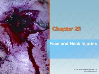
Chapter 25 Face and Neck Injuries
Introduction (1 of 2) • Face and neck are vulnerable to injury • Relatively unprotected positions on body • Soft-tissue injuries and fractures are common and vary in severity. • Some injuries are life-threatening. • Penetrating trauma to the neck may cause severe bleeding. • Open injury may result in an air embolism.
Introduction (2 of 2) • With appropriate prehospital and hospital care, a patient with a seemingly devastating injury can have a surprisingly good outcome.
The Head (1 of 2) • Cranium • Also referred to as the skull • Contains the brain • Most posterior portion is called the occiput. • Lateral portions on each side are called temples or temporal regions. • Forehead is called the frontal region.
The Head (2 of 2) • Cranium (cont’d) • Anterior to the ear, in the temporal region, you can feel the pulse of the superficial temporal artery.
The Face (1 of 6) • Composed of: • Eyes • Ears • Nose • Mouth • Cheeks • Jowls
The Face (2 of 6) • Six major bones include: • Nasal bone • Two zygomas • Two maxillae • Mandible
The Face (3 of 6) • The orbit of the eye is composed of: • Lower edge of the frontal bone of the skull • Zygoma • Maxilla • Nasal bone • Protects the eye from injury
The Face (4 of 6) • Only the proximal third of the nose is formed by bone. • The remaining two thirds are composed of cartilage.
The Face (5 of 6) • The exposed portion of the ear is composed entirely of cartilage covered by skin. • Pinna • Tragus • Superficial temporal artery
The Face (6 of 6) • About 1″ posterior to the external opening of the ear is the mastoid process. • The mandible forms the jaw and chin. • Motion of the mandible occurs at the temporomandibular joint.
The Neck (1 of 4) • Contains many important structures • Supported by the cervical spine • The upper part of the esophagus and the trachea lie in the midline of the neck. • The carotid arteries are found on either side of the trachea.
The Neck (2 of 4) • The larynx • Adam’s apple is located in the center of the neck. • Other portion of the larynx is the cricoid cartilage.
The Neck (3 of 4) • The larynx (cont’d) • The cricothyroid membrane lies between the thyroid cartilage and the cricoid cartilage. • Soft depression in the midline of the neck
The Neck (4 of 4) • The trachea • Below the larynx in the anterior midline of the neck • Connects the oropharynx and larynx with the main passages of the lungs • Sternocleidomastoid muscles • Originate from the mastoid process • Allow movement of the head
The Eye (1 of 7) • Globe-shaped, approximately 1″ in diameter • Located within a bony socket in the skull called the orbit • The orbit protects over 80% of the eyeball.
The Eye (3 of 7) • Clear, jellylike fluid near the back of the eye is called vitreous humor. • In front of the lens is a fluid called the aqueous humor, which can leak out in penetrating injuries.
The Eye (4 of 7) • The conjunctiva is a membrane that covers the eye. • The lacrimal glands produce fluid to keep the eye moist.
The Eye (5 of 7) • The sclera is the white, fibrous tissue that helps maintain the globular shape. • On the front of the eye, the sclera is replaced by a clear, transparent membrane called the cornea. • Allows light to enter the eye • The iris is a circular muscle behind the cornea.
The Eye (6 of 7) • The pupil is the opening in the center of the iris. • Allows light to move to the back of the eye • Anisocoria is a condition in which a person is born with different-sized pupils. • The lens lies behind the iris. • Focuses images on the retina at the back of the globe
The Eye (7 of 7) • The retina contains nerve endings. • Respond to light by transmitting nerve impulses through the optic nerve to the brain • The retina is nourished by a layer of blood vessels called the choroid. • Retinal detachment causes blindness.
Injuries of the Face and Neck (1 of 2) • Partial or complete obstruction of the upper airway may be the result. • Several factors may contribute. • Blood clots from heavy facial bleeding • Direct injuries to the nose and mouth, larynx, and trachea • Dislodgment of teeth or dentures in the throat
Injuries of the Face and Neck (2 of 2) • Several factors (cont’d) • Swelling that accompanies direct and indirect injury • Airway may be affected when the patient’s head is turned to the side • Possible injuries to the brain and/or cervical spine
Soft-Tissue Injuries • Very common • Face and neck are extremely vascular • Swelling may be more severe. • Skin and tissues in these areas have a rich blood supply. • A blunt injury can cause a hematoma. • Source: Courtesy of Rhonda Beck
Dental Injuries (1 of 2) • Mandible injuries are common. • Most of these injuries are the result of vehicle collisions and assaults. • Signs of mandible fractures include: • Misalignment of the teeth • Numbness of the chin • An inability to open the mouth
Dental Injuries (2 of 2) • Maxillary fractures are usually found after blunt force high-energy impacts. • Signs of maxillary fractures include: • Massive facial swelling • Instability of the facial bones • Misalignment of teeth • Fractured and avulsed teeth are common following facial trauma.
Patient Assessment • Patient assessment steps • Scene size-up • Primary assessment • History taking • Secondary assessment • Reassessment
Scene Size-up (1 of 2) • Scene safety • Observe for hazards and threats. • Assess for potential violence and environmental hazards. • Eye protection and face mask are standard. • Carry several pairs of gloves. • Determine the number of patients.
Scene Size-up (2 of 2) • Mechanism of injury/nature of illness • Assess the scene. • Common MOI for face and neck injuries: • Motor vehicle accidents • Sports • Falls • Penetrating trauma • Blunt trauma
Primary Assessment (1 of 7) • Focuses on identifying and managing life-threatening concerns • Perform a rapid scan. • Form a general impression. • Look for important indicators about the seriousness of the patient’s condition. • Injuries may be very obvious, or hidden.
Primary Assessment (2 of 7) • Form a general impression (cont’d). • Control blood loss with direct pressure. • Consider the need for manual spinal stabilization. • Check for responsiveness using the AVPU scale.
Primary Assessment (3 of 7) • Airway and breathing • Ensure a clear and patent airway. • If the patient is unresponsive, consider a properly sized oropharyngeal airway. • Palpate the chest wall for DCAP-BTLS. • Face and throat injuries increase the need for airway and breathing maintenance.
Primary Assessment (4 of 7) • Circulation • Quickly assess pulse rate and quality. • Determine skin condition, color, and temperature. • Check capillary refill time. • Significant bleeding is an immediate life threat.
Primary Assessment (5 of 7) • Transport decision • Patients with airway or breathing problems or with significant bleeding need to be transported immediately. • A patient with internal bleeding must be transported quickly for treatment by a physician.
Primary Assessment (6 of 7) • Transport decision (cont’d) • Signs of hypoperfusion include: • Tachycardia • Tachypnea • Low blood pressure • Weak pulse • Cool, moist, pale skin
Primary Assessment (7 of 7) • Transport decision (cont’d) • Even if the patient has no signs of hypoperfusion, there is the possibility of eye injuries. • The patient should be transported rapidly. • Surgery will need to be accomplished within 30 minutes or permanent blindness may result.
History Taking • Investigate the chief complaint. • Obtain a medical history. • Be alert for injury-specific signs and symptoms. • Be aware of pertinent negatives. • Gather a SAMPLE history from the patient, or from friends and family.
Secondary Assessment (1 of 4) • Physical examinations • If multiple systems have been affected, start with a full-body scan looking for DCAP-BTLS. • Do not delay transport to complete a thorough physical exam. • Focus on the isolated injury, the patient’s complaint, and the body region affected.
Secondary Assessment (2 of 4) • Physical examinations (cont’d) • Ensure that control of bleeding is maintained and note injury location. • Inspect the open wound for any foreign matter or impaled object. • Use both your eyes and your hands. • Assess all underlying systems.
Secondary Assessment (3 of 4) • Physical examinations (cont’d) • When evaluating the eyes, start with the outer aspect and work toward the pupils. • Look for discoloration, clarity of vision, bleeding, redness, eye symmetry, and pupil size and reaction to light. • Brain injury, nerve disease, glaucoma, and meningitis are causes of unequal pupils.
Secondary Assessment (4 of 4) • Vital signs • Assess vital signs to obtain a baseline. • You must be concerned with visible bleeding and unseen bleeding inside a body cavity. • With facial and throat injuries, baseline information is very important. • Use appropriate monitoring devices.
Reassessment (1 of 4) • Repeat the primary assessment. • Reassess vital signs and the chief complaint. • Reassess the patient’s condition every 5 minutes. • Interventions • Provide complete spinal immobilization if necessary.
Reassessment (2 of 4) • Interventions (cont’d) • Maintain an open airway, be prepared to suction, and consider an oropharyngeal airway. • Whenever you suspect significant bleeding, provide high-flow oxygen. • Control visible bleeding.
Reassessment (3 of 4) • Interventions (cont’d) • If the patient has signs of hypoperfusion, treat aggressively for shock and provide rapid transport. • Communication and documentation • Include a description of the MOI and the position in which you found the patient. • Document the method used to remove the patient from the vehicle.
Reassessment (4 of 4) • Communication and documentation (cont’d) • Recognize, estimate, and report the amount of blood loss. • Inform the hospital about all injuries involving the head and neck.
Emergency Medical Care (1 of 5) • Treat soft-tissue injuries to the face and neck the same as soft-tissue injuries elsewhere on the body. • Assess ABCs and life threats first. • Open and clear the airway. • Avoid moving the neck in patients with suspected cervical spine injuries.
Emergency Medical Care (2 of 5) • Control bleeding by applying direct manual pressure with a dry, sterile dressing. • Use roller gauze, wrapped around the head, to hold a pressure dressing in place. • Do not apply excessive pressure if an underlying skull fracture is suspected.
Emergency Medical Care (3 of 5) • Apply ice locally to injuries that do not break the skin. • For soft-tissue injuries around the mouth, check for bleeding inside the mouth. • Broken teeth and tongue lacerations may cause extensive bleeding and obstruction of the upper airway.
Emergency Medical Care (4 of 5) • Physicians can sometimes graft a piece of avulsed skin back into position. • If you find portions of avulsed skin: • Wrap in a sterile dressing. • Place in a plastic bag. • Keep cool, but do not place directly on ice. • Label and deliver to the emergency department.