Download
1 / 77
770 likes | 802 Views
Explore the essential role of blood proteins in maintaining colloid osmotic pressure, pH balance, and electrolyte levels, and learn how they contribute to the intricate processes within the bloodstream. Gain insights into protein structures and separation techniques for detailed analysis.

E N D
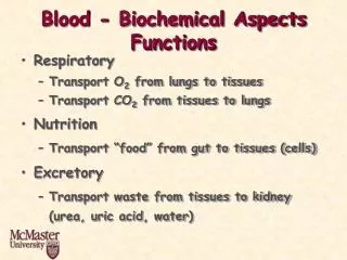
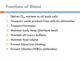
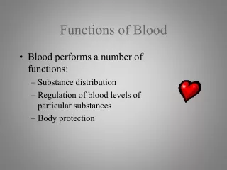
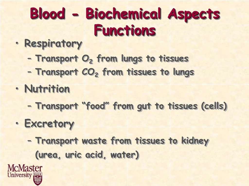
Blood - Biochemical AspectsFunctions • Respiratory • Transport O2 from lungs to tissues • Transport CO2 from tissues to lungs • Nutrition • Transport “food” from gut to tissues (cells) • Excretory • Transport waste from tissues to kidney (urea, uric acid, water)
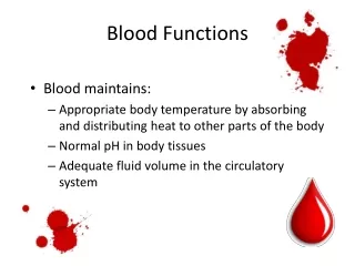
Regulatory • Water Content of Tissues • Water exchanged through vessel walls to tissue (interstitial fluid) • Body Temperature • Water- high heat capacity, thermal conductivity, heat of vaporization • Typical heat generation is 3000 kcal/day • Protective • Antibodies, antitoxins, white blood cells (WBC)
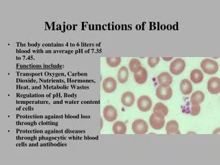
Blood composition • 5-6 L in an adult • 70 mL/kg of body weight • Suspension of cells in a carrier fluid (plasma) • Cells - 45% by volume • Plasma - 55% by volume • Cells • Red cells (erythrocytes) • 5x10⁶/mL • White cells (leukocytes) • 7x10³/mL • Platelets (thrombocytes) • 3x10⁵/mL
Plasma composition • Water - 90% of plasma volume • Proteins - 7% of plasma volume • Inorganic - 1% of plasma volume • Na+, K+, Mg2+, Ca2+, PO43-… • Organic - 2% of plasma volume • urea, fats, cholesterol, glucose ...
Male versus female • Hematocrit (% volume that is red cells) • 40-50% in males • 35-45% in females
ProteinsSee Lehninger Chapter 3-6 • Proteins are polyamino acids • Macromolecules - MW 5000 - several million • Insulin - MW = 6000 • Hemoglobin - MW = 68 000
O O ~NHCHC-NHCHC~ R1 R2 Peptide bond R NH2CH COOH Amino Acid Structure Protein Structure
20 common amino acids (AA) • Classified based on the properties of the R groups Acidic Glutamic Acid Basic Lysine
Polar Tyrosine Apolar Glycine
Amino Acids and Proteins • Acidic and basic groups are charged at blood / physiologic pH • Proteins are polyelectrolytes • pH of zero net charge (pI or isoelectric point) depends on amino acid composition of protein • Blood proteins negative at pH 7.4 • more COO- than NH3+, pI < 7.4
pI • Protein has many negative charges • Requires H+ to neutralize • Therefore low pI • Consider a protein with pI = 4 • If pH increases above pI protein becomes? • If pH decreases below pI protein becomes? • Higher the pI the more +,- is protein?
Need to go to a higher pH to neutralize or compensate for + charges • Minimum solubility occurs at pI since there is no intermolecular repulsion • At pH 7.4 (blood pH), all blood proteins are negative and therefore have pI’s less than 7.4
Protein Structure • Four levels • Primary structure: sequence of amino acids • 20 amino acids in long chain molecules • many possible combinations • Secondary structure: arrangement of the chains in space (conformation of chains) • a-helix: coil shape (due to H bonding) • b-sheet: stretched zig-zag peptide chain (H bonding • random coil: similar to synthetic polymers
Tertiary structure: folding of chains into 3 dimensional shape due to H bonding, S-S bonds and hydrophobic interactions • Several different types of secondary structure within the full three dimensional structure of a large protein • Quaternary structure: present in proteins with several polypeptide chains, arrangement and interelationship of the chains due to S-S bridging • Four levels result in well defined shape and chemical structure essential for function of protein
Plasma Proteins • More than 200 • Most abundant • Albumin - 4-5 g/100 mL • g-glubulins - ~1 g/100 mL • fibrinogen - 0.2-0.4g/100 mL • Original classification by zone electrophoresis at pH 8.6 • Separation by pI with several molecular weight species within each group
Zone Electrophoresis of Plasma Proteins + - globulins albumin g b a1 a2 pI 6.0 5.6 5.1 4.7
Protein Separation • Size Exclusion Chromatography (SEC) • Porous matrix (sephadex)
Affinity chromatography • molecule attached to a column that specifically binds the protein of interest • Coenzyme / enzyme • Antigen / Antibody
SDS-PAGE (polyacrylamide gel electrophoresis) • Separates by size • Proteins are complexed with SDS to give the same charge density
Two Dimensional Electrophoresis Decreasing Mr Decreasing pI
Functions of Plasma Proteins • Maintenance of: • Colloid osmotic pressure (p) • pH • electrolyte balance • COP relates to blood volume DP = p Protein sol’n Water
If membrane present p important • “Isotonic” - same osmotic pressure • Human blood - 300 milliOsmoles /L • Normal saline - 0.9% NaCl by weight • 0.15 mol/L • 0.30 mol/L of particles • Calculate osmotic pressure from concentration?
By analogy with the ideal gas law • In blood, which protein contributes most to p? • Low molecular weight, high concentration
Colloid - large particle that cannot easily cross a membrane • Stays in the compartment • In blood pprotein = 20-30 mmHg • Total ~ 5000 mmHg • Protein stays in the blood as p is maintained in the blood • Water content is therefore maintained
H2O Hb • Hypotonic - lower p than normal • Hemolysis of RBC H2O Ghost Cells • Hypertonic - higher p than normal • Hemolysis of RBC Crenated Cells Hypertonic 1.5% NaCl
Functions of Plasma Proteins (cont’d) • Transport of ions, fatty acids, steroids, hormones etc. • Albumin (fatty acids), ceruloplasmin (Cu2+), transferrin (Fe), lipoproteins (LDL, HDL) • Nutritional source of amino acids for tissues • Hemostasis (coagulation proteins) • Prevention of thrombosis (anticoagulant proteins) • Defense against infection (antibodies, complement proteins)
Function and Properties of Selected Plasma Proteins • Consider three abundant plasma proteins • Structure, function • Coagulation, fibrinolysis, complement
Albumin • MW 66 000 • Single chain, 580 amino acids, sequence is known • Dimensions - Heart shaped molecule • 50% a helix[He and Carter, Nature, 358 209 (1992)] • Modeled as: 80 Å 30 Å
Synthesis • Mainly liver cells then exported • Assembly time on ribosome ~ 1-2 min • t0.5 in circulation - 19 days • 14 g lost per day • 0.4 mg synthesized per hour per g of liver • Need liver of approximately 1.5 kg in weight to maintain
Functions • “Colloid” osmotic pressure of blood is 80% due to albumin • relatively low molecular weight • regulates water distribution • Transport of fatty acids • Liver to tissues, binding • Source of amino acids for tissue cells (pinocytosis) • 60% albumin in tissue (interstitial) fluid
g-Globulins • 20% of plasma proteins • “g” refers to electrophoretic mobility • Represents a group of proteins of variable structure • immunoglobulins • Main functional task is immunochemical • Antibodies - combine with specific antigens
Basic 4 chain structural unit • MW = 2x55000 +2x27000 = 160000
Variable region varies with respect to primary, secondary and tertiary structures • Basis of specificity of antigen binding (106 average number) • 5 classes of immunoglobulins • IgG, IgA, IgM, IgD, IgE • Different structures of constant regions of heavy chains • Some are polymers (multiples of 4 chain unit - IgA - dimer - MW 350 000, IgM - pentamer - MW 900 000 • See any immunology book for more details
Functions • Primary function is antigen binding (immune response) • Secondary function is complement binding (after antigen) • Each class has different functions • IgE - allergic reactions (defence) • IgA - secretory protein, high concentration in external fluids (saliva, tears) • IgD - ? Involved in differentiation of B lymphocytes (found on the surface of B-lymphocytes)
Synthesis • In lymphocytes (T and B) • Made in response to presence of antigen (“foreign” macromolecule, virus particle etc.)
Fibrinogen • Coagulation • Structure • MW 340 000 • Sequence of amino acids is known (3000) • 4y, 3y structure • 6 polypeptide chains, 2a (67,000), 2b (56,000), 2g (47,000)
a b g disulfide Triple dumbell model (EM) 450 Å 90 Å D E D a’s, b’s and g’s are intertwined
Thrombin Fibrinogen Fibrin Plasmin Fibrin Degradation (FDP) • Function • Blood coagulation (clotting) • Plasmin is end product of fibrinolytic system • Clot needs to be removed • Not needed forever • Could embolize to lungs, brain
Sickle Cell Anemia • Occurs because of a minor variation in one amino acid in the b chain of Hb • Results in Hb that, when exposed to low O2 concentrations precipitates into long crystals • Elongate cell • Damage cell membrane • Decrease in amount of RBC
Cellular Elements of Blood • Red cells • 40 - 50% of blood volume • 5 x 10⁶ cells /mL • “bag” of hemoglobin • non-nucleated • no proliferation • cell membrane in excess so that deformation does not rupture • Shape • Biconcave disc • 8 mm in diameter, 2.7 mm thick, volume ~ 90 mm3, area ~ 160 mm2
Why this shape? • Area to volume ratio is high (maximal?) • Facilitates diffusion of O2 and CO2 • minimal distance of contents from surface • Originates in bone marrow (hematopoiesis) • Molecular explanation based on the properties of the proteins in the cell membrane is found in Elgsaeter et al. Science, 234, 1217 (1986)
Oxygen Binding of Hb • Blood must carry 600 L of O2 from lungs to tissues each day • Very little carried in plasma since O2 only sparingly soluble • Nearly all bound and transported by Hb of RBC • Possible for Hb to carry four O2 molecules, one on each a chain, one on each b chain
O2 depleted Hb solution placed in contact with O2(g) • Equilibrium reaction • Fraction (s) of Hb converted to oxyhemoglobin