Download
1 / 12
130 likes | 426 Views
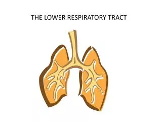
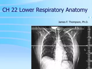
CH 22 Lower Respiratory Anatomy. James F. Thompson, Ph.D. The Lungs. apex. Apex, base Left lung has cardiac notch R lung thicker, broader, shorter than L. cardiac notch. base. The Lobes of the Lungs. Lobes are separated by fissures

E N D
CH 22 Lower Respiratory Anatomy James F. Thompson, Ph.D.
The Lungs apex • Apex, base • Left lung has cardiac notch • R lung thicker, broader, shorter than L cardiac notch base
The Lobes of the Lungs Lobes are separated by fissures • Each lobe has a secondarybronchus and tertiary bronchi • 10 tertiary bronchi/lung • tertiary bronchi supply each bronchopulmonary (BP) segment • Tumors, infections or abscesses initially localized
The Lobules of the Lungs • Each bronchopulmonary segment has elastic connective tissue around a lymphatic vessel, an arteriole, a venule, and the terminal bronchiole • Terminal bronchiole continues to branch • Respiratory bronchiole Alveolar duct Alveolar sac Alveoli
The Alveoli of the Lungs • Alveolar ducts open into alveolar sacs & alveoli • Alveolar sacs - 2 or more alveoli • Thin walls • simple squamous epithelium • thin elastic basement membrane • capillary endothelium • ~300 million alveoli • Total surface area 1/2 to 2/3’s of a tennis court covered by ~1 liter of blood!
The Alveolus • 2 cell types in walls • type I alveolar pulmonary epithelial (squamous) cells • Make ACE • type II alveolaror septal cells • secrete surfactant alveolar fluid • (detergent-like substance) • Alveolar macrophages (“dust cells”) patrol the alveolar walls
The Alveolar Wall • Capillaries (endothelial cells) surround alveoli for gas exchange • Smooth muscle controls airway resistance Alveolar-capillary (respiratory) interface
The Alveolar Wall Alveolar-capillary (respiratory) interface • Interface consists of: • Alveolar epithelium • Epithelial basement membrane • Capillary basement membrane • Endothelial cells of capillary O2 CO2 • Total thickness • 0.5 µm • short diffusion distance
The Alveolar Space • Alveolar fluid • Surface tension • Attraction of water to other water molecules • Surfactant: phospholipids decrease surface tension • Respiratory distress syndrome
Blood Supply for the Resp. System • Pulmonary circulation • blood for gas exchange • low pressure system - 25/6 mm Hg • Bronchial (systemic) circulation • a small supply from the aorta for nutrients and O2 • supplies entire lung (except alveoli) • high pressure system • bronchial arteries to bronchial capillaries, then to both pulmonary veins and bronchial veins • Pulmonary plexus • Area of root where nerve fibers enter lung • Sympathetic/parasympathetic/visceral sensory
The Pleura • Lungs – housed in the bony thorax • Two pleural membranes with pleural cavity between • outer - parietal pleura • inner - visceral pleura • pleural cavity – lowered intraplural pressure is normal • Rib cage • protective and flexible