Download
1 / 26
260 likes | 352 Views
The study evaluates the diagnostic performance of FFRCT for identifying significant coronary artery disease compared to invasive FFR, focusing on intermediate stenoses. Using advanced imaging technology and patient-specific modeling, this research aims to enhance diagnostic precision in cardiac care.

E N D
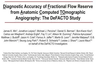
Diagnostic Accuracy of Fractional Flow Reserve from Anatomic Computed TOmographic Angiography: The DeFACTO Study James K. Min1; Jonathon Leipsic2; Michael J. Pencina3; Daniel S. Berman1; Bon-Kwon Koo4; Carlos van Mieghem5; Andrejs Erglis6; Fay Y. Lin7; Allison M. Dunning7; Patricia Apruzzese3; Matthew J. Budoff8; Jason H. Cole9; Farouc A. Jaffer10; Martin B. Leon11; Jennifer Malpeso8; G.B. John Mancini12; Seung-Jung Park13, Robert S. Schwartz14; Leslee J. Shaw15, Laura Mauri16 on behalf of the DeFACTO Investigators 1Cedars-Sinai Heart Institute, Los Angeles, CA; 2St. Paul’s Hospital, Vancouver, British Columbia; 3Harvard Clinical Research Institute, Boston, MA; 4Seoul National University Hospital, Seoul, Korea; 5Cardiovascular Center, Aalst, Belgium; 6Pauls Stradins Clinical University Hospital, Riga, Latvia; 7Cornell Medical College, New York, NY; 8Harbor UCLA, Los Angeles, CA; 9Cardiology Associates, Mobile, AL; 10Massachusetts General Hospital, Harvard Medical School, Boston, MA; 11Columbia University Medical Center, New York, NY; 12Vancouver General Hospital, Vancouver, British Columbia; 13Asan Medical Center, Seoul, Korea; 14Minneapolis Heart Institute, Minneapolis, MN; 15Emory University School of Medicine, Atlanta, GA; 16Brigham and Women’s Hospital, Boston, MA
Disclosures • Study funding provided by HeartFlow which had no involvement in the data analysis, abstract or manuscript preparation • No study investigator had any financial interest related to the study sponsor
Background • Coronary CT Angiography: • High diagnostic accuracy for anatomic stenosis • Cannot determine physiologic significance of lesions1 • Fractional Flow Reserve (FFR): • Gold standard for diagnosis of lesion-specific ischemia2 • Use improves event-free survival and cost effectiveness3,4 • FFR Computed from CT (FFRCT): • Novel non-invasive method for determining lesion-specific ischemia5 1Min et al. J Am Coll Cardiol 2010; 55: 957-65; 2Piljs et al. Cath Cardiovasc Interv 2000; 49: 1-16; 3Tonino et al. N Engl J Med 2009; 360: 213-24; 4Berger et al. J Am Coll Cardiol 2005; 46: 438-42; 5Kim et al. Ann Biomed Eng 2010; 38: 3195-209
Overall Objective • To determine the diagnostic performance of FFRCT for detection and exclusion of hemodynamically significant CAD
Study Endpoints • Primary Endpoint: Per-patient diagnostic accuracy of FFRCT plus CT to diagnose hemodynamically significant CAD, compared to invasive FFR reference standard • Null hypothesis rejected if lower bound of 95% CI < 0.70 • 0.70 represents 15% increase in diagnostic accuracy over myocardial perfusion imaging and stress echocardiography, as compared to FFR1 • 252 patients: >95% power • Secondary Endpoint: • Diagnostic performance for intermediate stenoses (30-70%) 1Mellikan N et al. JACC: Cardiovasc Inter 2010, 3: 307-314; 2Jung PH et al. Eur Heart J 2008; 29: 2536-43
Study Criteria Inclusion Criteria: • Underwent >64-row CT • Scheduled for ICA within 60 days of CT • No intervening cardiac event Exclusion Criteria: • Prior CABG • Suspected in-stent restenosis • Suspected ACS • Recent MI within 40 days of CT ICA = Invasive coronary angiography; CABG = coronary artery bypass surgery; ACS = acute coronary syndrome; MI = myocardial infarction
Study Procedures • Intention-to-Diagnose Analysis • Independent blinded core laboratories for CT, QCA, FFR and FFRCT • FFRCT for all CTs received from CT Core Laboratory • CT: Stenosis severity range1 • 0%, 1-24%, 25-49%, 50-69%, >70-89%, >90% • QCA: Stenosis severity (%) • FFR: At maximum hyperemia during ICA • Definition: (Mean distal coronary pressure) / (Mean aortic pressure) • Obstructive CAD: >50%stenosis (CT and QCA) • Lesion-Specific Ischemia: <0.80 (FFR and FFRCT)2 1Raff GL et al. J Cardiovasc Comp Tomogr 2009; 3: 122-36; 2Tonino PA et al. N Engl J Med 2009; 360: 213-24; FFR, subtotal / total occlusions assigned value of 0.50; FFRCT, subtotal / total occlusions assigned value of 0.50, <30% DS assigned value of 0.90
Study Procedures: FFRCT FFRCT: Derived from typical CT • No modification to imaging protocols • No additional image acquisition • No additional radiation • No administration of adenosine • Selectable at any point of coronary tree Patient-Specific Coronary Pressure: • Image-based modeling • Heart-Vessel Interactions • Physiologic conditions, incl. Hyperemia • Computational fluid dynamics to calculate FFRCT Simulation of coronary pressure and flow
Patient Enrollment • Study Period • October 2010 – 2011 • Study Sites • 17 centers from 5 countries • Study Enrollment (n=285) • n=33 excluded • Final study population • Patients (n=252) • Vessels (n=406)
Patient and Lesion Characteristics • ICA • Stenosis >50% 47% • Mean Stenosis 47% • FFR • FFR < 0.80 37% • CT • Stenosis >50% 53% • Calcium Score 381 • Location • LAD 55% • LCx 22% • RCA 23% Abbreviations: MI = myocardial infarction; PCI = percutaneous intervention; FH = family history; CAD = coronary artery disease; FFR = fractional flow reserve; CACS = coronary artery calcium score; LAD = left anterior descending artery; LCx = left circumflex artery; RCA = right coronary artery *N=408 vessels from 252 patients; ^N=406 vessels from 252 patients
Per-Patient Diagnostic Performance FFRCT <0.80 CT >50% % N=252 95% CI FFRCT CT 95% CI 67-78 58-70 95% CI 84-95 77-90 95% CI 46-83 34-51 95% CI 60-74 53-67 95% CI 74-90 61-81
Discrimination Per-Patient Per-Vessel • Greater discriminatory power for FFRCT versus CT stenosis • Per-patient (Δ 0.13, p<0.001) • Per-vessel (Δ 0.06, p<0.001) AUC AUC FFRCT 0.81 (95% CI 0.75, 0.86) CT 0.68 (95% CI 0.62, 0.74) FFRCT 0.81 (95% CI 0.76, 0.85) CT 0.75 (95% CI 0.71, 0.80) *AUC = Area under the receiving operating characteristics curve
Case Examples: Obstructive CAD FFRCT CT ICA and FFR Case 1 FFR 0.65 = Lesion-specific ischemia FFRCT 0.62 = Lesion-specific ischemia LAD stenosis FFRCT ICA and FFR CT Case 2 FFRCT 0.87 = No ischemia FFR 0.86 = No ischemia RCA stenosis
Per-Patient Diagnostic Performance for Intermediate Stenoses by CT (30-70%) FFRCT <0.80 CT >50% N=83 95% CI 61-80 63-92 95% CI 63-92 53-77 95% CI 53-77 53-77 95% CI 39-68 20-53 95% CI 75-95 55-79 95% CI FFRCT CT
Case Example: Intermediate Stenosis CT Core Lab 31-49% stenosis QCA Core Lab 50-69% stenosis CT ICA and FFR FFRCT FFR 0.74 FFRCT 0.71 FFRCT 0.71 = Lesion-specific ischemia RCA intermediate stenosis FFR 0.74 = Lesion-specific ischemia
Limitations • ICA was performed based upon CT results (referral bias) • Did not interrogate every vessel with invasive FFR • Did not solely enroll patients with intermediate stenosis1,2 • Did not test whether FFRCT-based revascularization reduces ischemia3 • Did not enroll prior CABG / In-Stent Restenosis / Recent MI 1Koo BK et al. 2012 EuroPCR Scientific Sessions, 2Fearon et al. Am J Cardiol 2000: 86: 1013-4; 2Melikian N et al. JACC Cardiovasc Interv 2010; 3: 307-14
Conclusions • FFRCT demonstrated improved accuracy over CT for diagnosis of patients and vessels with ischemia • FFRCT diagnostic accuracy 73% (95% CI 67-78%) • Pre-specified primary endpoint >70% lower bound of 95% CI • Increased discriminatory power • FFRCT superior to CT for intermediate stenoses • FFRCT computed without additional radiation or imaging • First large-scale demonstration of patient-specific computational models to calculate physiologic pressure and velocity fields from CT images • Proof of feasibility of FFRCT for diagnosis of lesion-specific ischemia
Patient-Specific Computation of FFRCT (2) (3) (6) (1) (4) (5) • Image-Based Modeling – Segmentation of patient-specific arterial geometry • Heart-Vessel Interactions – Allometric scaling laws relate caliber to pressure and flow • Microcirculatory resistance – Mophometry laws relate coronary dimension to resistance • Left Ventricular Mass – Lumped-parameter model couples pulsatile coronary flow to time-varying myocardial pressure • Physiologic Conditions – Blood as Newtonian fluid adjusted to patient-specific viscosity • Induction of Hyperemia – Compute maximal coronary vasodilation • Fluid Dynamics – Navier-Stokes equations applied for coronary pressure 140 mcg/kg/min
Diagnostic Accuracy of Fractional Flow Reserve from Anatomic Computed TOmographic Angiography: The DeFACTO Study James K. Min1; Jonathon Leipsic2; Michael J. Pencina3; Daniel S. Berman1; Bon-Kwon Koo4; Carlos van Mieghem5; Andrejs Erglis6; Fay Y. Lin7; Allison M. Dunning7; Patricia Apruzzese3; Matthew J. Budoff8; Jason H. Cole9; Farouc A. Jaffer10; Martin B. Leon11; Jennifer Malpeso8; G.B. John Mancini12; Seung-Jung Park13, Robert S. Schwartz14; Leslee J. Shaw15, Laura Mauri16 on behalf of the DeFACTO Investigators 1Cedars-Sinai Heart Institute, Los Angeles, CA; 2St. Paul’s Hospital, Vancouver, British Columbia; 3Harvard Clinical Research Institute, Boston, MA; 4Seoul National University Hospital, Seoul, Korea; 5Cardiovascular Center, Aalst, Belgium; 6Pauls Stradins Clinical University Hospital, Riga, Latvia; 7Cornell Medical College, New York, NY; 8Harbor UCLA, Los Angeles, CA; 9Cardiology Associates, Mobile, AL; 10Massachusetts General Hospital, Harvard Medical School, Boston, MA; 11Columbia University Medical Center, New York, NY; 12Vancouver General Hospital, Vancouver, British Columbia; 13Asan Medical Center, Seoul, Korea; 14Minneapolis Heart Institute, Minneapolis, MN; 15Emory University School of Medicine, Atlanta, GA; 16Brigham and Women’s Hospital, Boston, MA
The DeFACTO Study: Background • Coronary CT Angiography: • High diagnostic accuracy for anatomic stenosis • Cannot determine physiologic significance of lesions1 • Fractional Flow Reserve (FFR): • Gold standard for diagnosis of lesion-specific ischemia2 • Use improves event-free survival and cost effectiveness3,4 • FFR Computed from CT (FFRCT): • Novel non-invasive method for determining lesion-specific ischemia5,6 1Min et al. J Am Coll Cardiol 2010; 55: 957-65; 2Piljs et al. Cath Cardiovasc Interv 2000; 49: 1-16; 3Tonino et al. N Engl J Med 2009; 360: 213-24; 4Fearon WF et al. Circulation 2010; 122: 2545-50; 5Kim et al. Ann Biomed Eng 2010; 38: 3195-209; 6Koo BK et al. J Am Coll Cardiol 2011; 58: 1989-97.
The DeFACTOStudy: Patient Enrollment • Study Period • October 2010 – 2011 • Study Sites • 17 centers from 5 countries • Study Enrollment (n=285) • n=33 excluded • Final study population • Patients (n=252) • Vessels (n=406)
The DeFACTO Study: Per-Patient Diagnostic Performance • Greater diagnostic accuracy of FFRCT versus CT stenosis • 9% absolute improvement in diagnostic accuracy • Improved discriminatory power for FFRCT versus CT stenosis • Per-patient (Δ 0.13, p<0.001)
The DeFACTO Study: Intermediate Stenoses (30-70%) FFRCT 0.71 = Lesion-specific ischemia of an intermediate stenosis (30-70%) - Concordant and in agreement with invasive FFR RCA intermediate stenosis FFR 0.74 = Lesion-specific ischemia
Conclusions • FFRCT demonstrated improved accuracy over CT for diagnosis of patients and vessels with ischemia • FFRCT diagnostic accuracy 73% (95% CI 67-78%) • Pre-specified primary endpoint >70% lower bound of 95% CI • Increased discriminatory power • FFRCT superior to CT for intermediate stenoses • FFRCT computed without additional radiation or imaging • First large-scale demonstration of patient-specific computational models to calculate physiologic pressure and velocity fields from CT images • Proof of feasibility of FFRCT