Download

1 / 26
260 likes | 562 Views
Health. Health. Environment. Lifestyle factors. Genetic. Poverty. Cultural. Pre existing disease. Statistics. 40% of pregnancies are not planned (BBC News 2004) Some texts state 50%1:4 pregnancies result in miscarriage15.1% of all stillbirths are from congenital abnormalities whilst 50% of stillbi

E N D
1. Preconception Care & Fertilisation, Embryology & Fetal Development Effective Midwifery practice
Level 6
Kate Quarrell
2. Health
3. Statistics 40% of pregnancies are not planned (BBC News 2004) Some texts state 50%
1:4 pregnancies result in miscarriage
15.1% of all stillbirths are from congenital abnormalities whilst 50% of stillbirths are unexplained (CEMACH 2006)
Neonatal deaths 48% are from immaturity and 22.4% from congenital abnormalities (CEMACH 2006)
4. Why Preconception care?
5. Pre conception care continued Example of smoking. Smoking in pregnancy has been positively linked to prematurity, low birth weight and stillbirth. Subsequently the baby is 15 times more at risk of sudden infant death syndrome.
Example of social factors. Infants born to families of socio-economic groups IV & V are twice as likely to die between the end of the first month of life and 1 year.
Example of maternal disease. Babies born to women with Type 1 or 2 diabetes have a 3.8 times higher perinatal mortality rate with congenital abnormality nearly twice that expected (CEMACH 2006 p 22)
6. Why study embryology? Knowledge is the key to un locking our minds to release us to provide the appropriate care.
What do we need to know about embryology?
Significant times in embryo development
Processes involved in that development
What can hinder healthy embryo and fetal development?
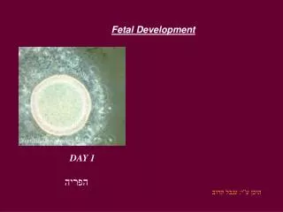
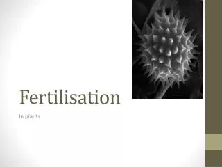
7. Significant landmarks - Fertilisation Formation of diploid zygote � single cell from fusion of haploid gametes.
Gametes formed by cell division known as meiosis which is a process by which the chromosome content is reduced to 23.
(Spermatogenesis or Oogenesis)
Process allows for mixing of maternal and paternal genes
8. Problems Spontaneous abortion
Genetic disorder. Anomalies of the base within the DNA so all cells affected e.g. Phenylketonuria.
50% associated with chromosomal abnormalities
More common in older women � probably associated with nondisjunction of chromosomes e.g. Down�s syndrome
9. Mitosis Mitotic cell division � formation of
Cells have identical genetic material
Totipotent � clones
Day 4 16-20 cells & is known as a Morula
10. Blastocyst Cells division becomes asymmetrical
Cells polarise
Loose their totipotency and begin to differentiate
Inner cell mass � fetus
Outer cell mass becomes the trophoblast which develops into the placenta
Implants day 6
11. Problems with Implantation Example Pelvic inflammatory disease, commonly develops between 15 -24 years. Ectopic pregnancy 1:10
Structural anomalies of the uterus might inhibit implantation
Intrauterine contraceptive disease
12. Cells 350 different types of human cells
Different functions
Cells process: division, differentiation, induction, migration & death
13. By day 14 From implantation of the blastocyst the inner cell mass is known as the embryo
Epiblast & Hypoblast = Bilaminar embryonic disc
Primitive streak develops � significance is the threshold at which experimentation must stop.
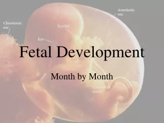
Amniotic & Chorionic cavities
14. Embryo 3-8 weeks Critical time for normal development
Particularly sensitive to external factors, environmental hazards, pharmacological agents, drug misuse
Organogenesis
Trilaminar disc folds into C shaped cylindrical embryo
Co-ordinated by genes � Homeobox
Cell differentiation
Tissue interaction & communication
Folding is due to different rate of growth
15. Gastrulation Cell migration and rearrangement � Week 3 Primitive streak in the midline
Bilaminar disc is converted into Trilaminar disc
3 Germ layers :Ectoderm, Mesoderm & Endoderm
2mm long
Notochord forms
Primitive heart
Mother first missed menstrual period
16. Trilaminar disc Ectoderm will form the epidermis & central nervous system
Mesoderm will form the bones, muscles and heart, blood vessels, kidneys and reproductive organs
Endoderm will form digestive tract, respiratory tract, glands & mucous membranes
17. Formation of the Neural tube - Neurulation Starts at 22- 23 days
Folding starts in the middle in both the cranial and caudal direction.
Cranial opening closes day 25 , caudal opening closes on day 27days
Folic acid is involved in DNA synthesis
Most women at this stage do no know they are pregnant
18. Development of the skeletal vertebral column Commences at week 4
Week 6 cartilaginous stage
Week 8 Ossification begins
19. Week 4 Heart begins to beat approximately 85 beats /minute
Outline of eyes
Upper limb buds
Lungs begin to form
Parts of gastro intestinal tract can be identified.
20. Week 8 Heart has 4 chambers
Upper limbs longer bent at the elbows
Fingers distinct but webbed
External genitalia still in sexless state but have begun to differentiate
By end of week 8 all body systems & organs are formed.
21. Summary of human development Growth � cell division
Morphogenesis � Development of form. Movements of sheets & masses of cells
Differentiation � Maturation of cells. Formation tissues and organs.
(Moore & Persuad 2003)
22. 8-12 Weeks Eye lids fuse
Fetal circulation functioning
Moves freely
Kidney�s function fetus passes urine ~10 weeks
Abdominal gut needs to be withdrawn into cavity by week 10
Ossification of bones begins 8 weeks
23. By 20 Weeks Most organs capable of functioning
Neurons formed between 10-18 weeks
Skin covered with vernix and lanugo
Brown fat deposited
Limbs are at mature proportions
Meconium present in gut
24. 24 Weeks Skin � thin, wrinkled, translucent & dark red
Lungs terminal sac phase (surfactant started to be produced 22weeks, increases significantly after 30weeks)
Sensory organs develop, fetus responds to noise
Length 32 cm
Weight 700g
Periods of sleep & activity
25. 28 Weeks Survival possible
Eyelids open
Length 37cm
Weight 1200g
Head circumference 26cm
Girl � small labia majora
Boy � scrotum � few rugae
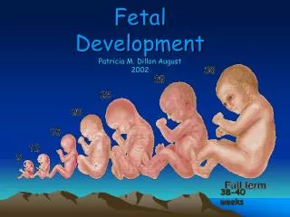
26. 32 Weeks Lanugo disappears from face
Ear cartilage soft
Lengths 43cm
Weight 2000g
Accumulation of fat
27. 36 Weeks Head circumference > abdominal circumference
Plantar creases visible
Head hair lengthens
Nails reach the tips of fingers
Lanugo vanishes from shoulder
Breast tissue nodule present 1-2 mm
Skin pale
Length 49cm
Head circumference 33cm
Weight 2900g